. 2020 Aug 25;117(34):20597-20606.
doi: 10.1073/pnas.2004445117. Epub 2020 Aug 11.

Atomistic structure and dynamics of the human MHC-I peptide-loading complex

Affiliations

Atomistic structure and dynamics of the human MHC-I peptide-loading complex

Olivier Fisette et al. Proc Natl Acad Sci U S A. .

Abstract

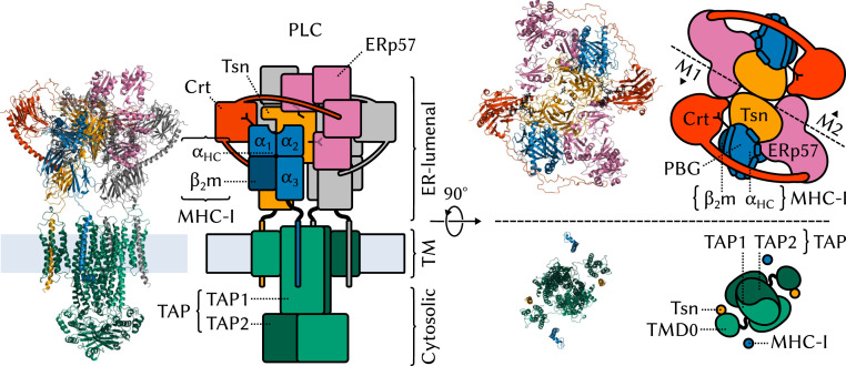
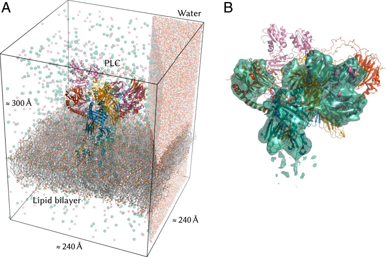
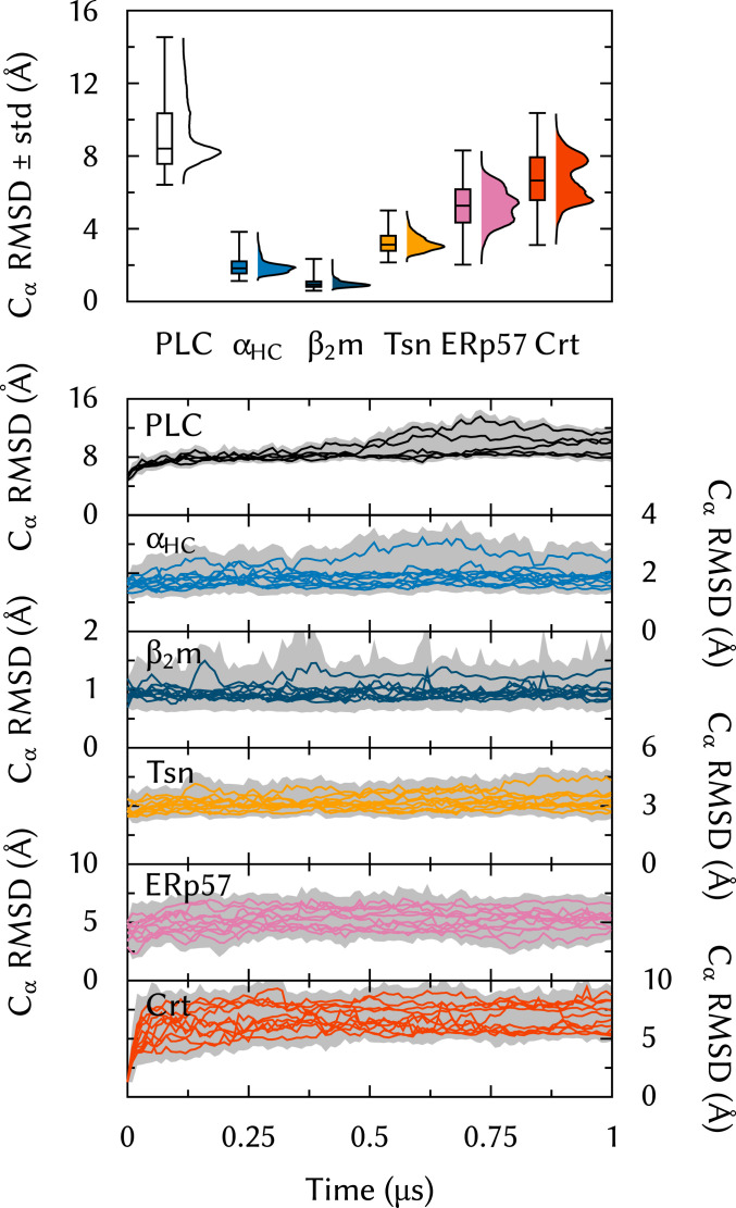
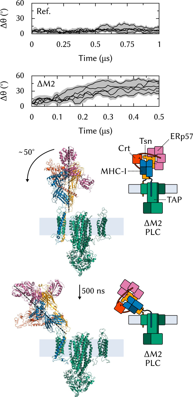
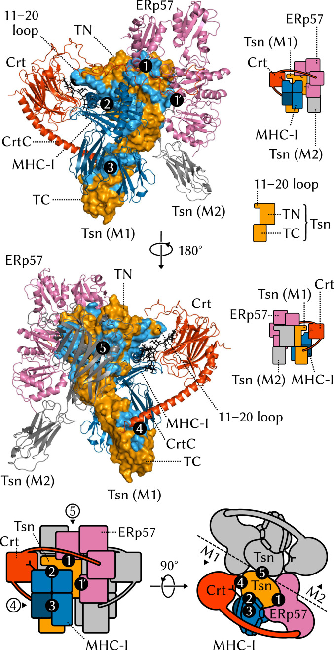
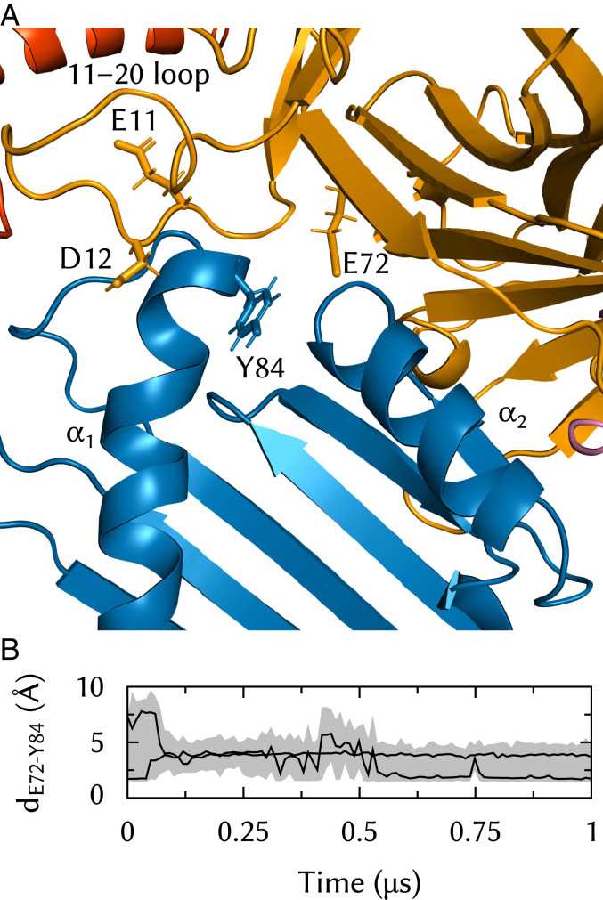
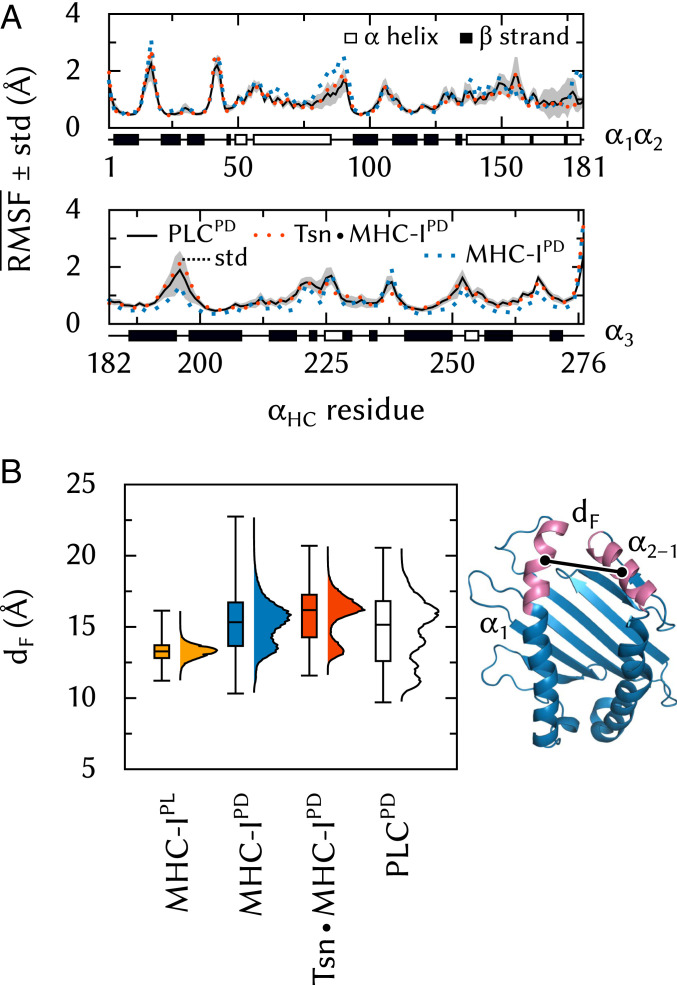
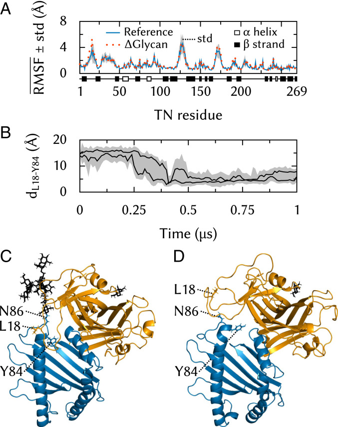
The major histocompatibility complex class-I (MHC-I) peptide-loading complex (PLC) is a cornerstone of the human adaptive immune system, being responsible for processing antigens that allow killer T cells to distinguish between healthy and compromised cells. Based on a recent low-resolution cryo-electron microscopy (cryo-EM) structure of this large membrane-bound protein complex, we report an atomistic model of the PLC and study its conformational dynamics on the multimicrosecond time scale using all-atom molecular dynamics (MD) simulations in an explicit lipid bilayer and water environment (1.6 million atoms in total). The PLC has a layered structure, with two editing modules forming a flexible protein belt surrounding a stable, catalytically active core. Tapasin plays a central role in the PLC, stabilizing the MHC-I binding groove in a conformation reminiscent of antigen-loaded MHC-I. The MHC-I-linked glycan steers a tapasin loop involved in peptide editing toward the binding groove. Tapasin conformational dynamics are also affected by calreticulin through a conformational selection mechanism that facilitates MHC-I recruitment into the complex.

Keywords: MHC-I; immunity; molecular dynamics simulations; peptide-loading complex; protein dynamics.


Conflict of interest statement

The authors declare no competing interest.

Figures

Fig. 1.
Fig. 1.
Structural overview of the MHC-I PLC. The PLC can be divided into three parts, ER-luminal, TM, and cytosolic. The ER-luminal part consists of two editing modules, M1 and M2, each of which contains one Tsn, ERp57, Crt, and MHC-I. MHC-I proteins (–3, 6, 7) are heterodimers formed of a variable α heavy chain (αHC) and an invariant, light β2 microglobulin (β2m). αHC comprises three soluble domains, two of which, α1 and α2, form the peptide-binding groove (PBG). MHC-Is have an N-linked branched glycan that reflects their loading status: A terminal glucose allows recognition and binding by Crt and acts as a signal that MHC-I should be recruited to the PLC for peptide editing; antigen-loaded MHC-Is that exit the ER are deglucosylated. Newly synthesized “empty” MHC-I proteins (eMHC-I) are unstable; in the PLC, Tsn acts as an MHC-I chaperone. Tsn (–10) is the central component of the editing modules. It has two ER-luminal domains: N-terminal TN and C-terminal TC. M1 and M2 are organized around a pseudosymmetry axis at the interface between the two Tsns. Tsn forms a complex with MHC-I and accelerates the off rate of low-affinity MHC-I–bound peptides to perform peptide editing (, – 13). Tsn is also disulfide bonded to ERp57 (14), a four-domain protein playing a structural role. Crt (15, 16) consists of three soluble domains, a globular lectin domain with a binding site for the monoglucosylated branch of the N-linked MHC-I glycan, a flexible P domain that extends over the PBG and contacts ERp57, and a calcium-sensing C domain with an extended α-helix that contacts Tsn. The transporter associated with antigen processing (TAP) (17) is the main component of both the TM and cytosolic parts of the PLC. TAP shuttles peptides from the cytosol to the ER, providing the PLC with its substrate for antigen processing. TAP is a heterodimer of TAP-1 and TAP-2, each of which has an N-terminal four-helix TM domain, TMD0, that provides a docking site for the TM helix of Tsn. A TM helix also anchors MHC-I to the ER membrane.
Fig. 2.
Fig. 2.
Atomistic model of the MHC-I peptide-loading complex. (A) The 1.6-million-atom molecular dynamics simulation system contains the complete human MHC-I PLC (75.939 atoms), embedded in a POPC lipid bilayer (101.036 atoms) and solvated by explicit water with Na+ and Cl ions. (Only a small slab of water is shown for clarity.) (B) The PLC model was built by fitting atomistic subunit structures to the cryo-EM density for a single editing module (EMD-3906) (5) and then duplicating this module and fitting to the cryo-EM data (EMD-3094) (5) for the pseudosymmetric assembly (cryo-EM density for a single module shown as a surface).
Fig. 3.
Fig. 3.
Cα rmsd from the starting structure, calculated for ER-luminal components individually and for the entire PLC.
Fig. 4.
Fig. 4.
Effects of ΔM2 on PLC structural stability. To measure the tilt of the editing module, θ is defined as the angle between the centers of geometry of the two soluble Tsn domains (TN and TC). Δθ is the deviation from the initial orientation; it is reported for the reference system (full PLC, Ref.) (Top) and for ΔM2 (Middle). (Bottom) The starting structure of ΔM2 and the structure with tilted editing module obtained after 500 ns of MD simulation.
Fig. 5.
Fig. 5.
Interfaces between ER-luminal Tsn domains and other PLC components. TM regions are not shown.
Fig. 6.
Fig. 6.
Interactions between Tsn E72 and the MHC-I binding groove in the vicinity of the F pocket. (A) E72 is lodged between MHC-I α1 and α2 and contacts Y84, which is also a suggested interaction partner of the Tsn 11–20 loop. E72 and residues E11 and D12 from that loop have been shown to be important for Tsn association with MHC-I (10). Only Tsn TN and MHC-I α1 and α2 are shown for clarity. (B) E72 to Y84 distance time series in two trajectories.
Fig. 7.
Fig. 7.
MHC-I dynamics in the PLC. (A) Effects of Tsn and PLC binding on RMSF along the αHC sequence. SD is shown for the PLC system. (B) Effects of peptide, Tsn, and PLC binding on binding-groove width dF, measured as the distance between the centers of mass of Cα atoms in residues 75 to 85 (α1) and 138 to 150 (α21).
Fig. 8.
Fig. 8.
Tsn 11–20 loop dynamics. (A) Effects of glycan removal on RMSF along the TN sequence. SD is shown for the reference system. (B) TN L18 to αHC Y84 distance time series. (C) Contact between the 11–20 loop and the MHC-I binding groove in a simulation of the full PLC. (D) Typical 11–20 loop conformation in a simulation of the ΔGlycan system.
Fig. 9.
Fig. 9.
Influence of the Crt C domain on Tsn TC conformational dynamics. (A) Tsn TC is lodged between MHC-I α3 and β2m and also contacted by the Crt C domain. (B) Cα rmsd of TC using TN Cα for structural superposition. (C) The average plane of the TC β-sheet region that contacts MHC-I (residues 273 to 277, 293 to 298, 327 to 329, 341 to 346) is used to measure TC rotation. (D) Angle θ of the β-sheet to its initial orientation, viewed from the top of the ER-luminal part of the PLC toward the membrane.

References

    1. Neefjes J., Jongsma M. L. M., Paul P., Bakke O., Towards a systems understanding of MHC class I and MHC class II antigen presentation. Nat. Rev. Immunol. 11, 823–836 (2011). - PubMed
    1. Blum J. S., Wearsch P. A., Cresswell P., Pathways of antigen processing. Annu. Rev. Immunol. 31, 443–473 (2013). - PMC - PubMed
    1. Rock K. L., Reits E., Neefjes J., Present yourself! by MHC class I and MHC class II molecules. Trends Immunol. 37, 724–737 (2016). - PMC - PubMed
    1. Hulpke S., Tampé R., The MHC I loading complex: A multitasking machinery in adaptive immunity. Trends Biochem. Sci. 38, 412–420 (2013). - PubMed
    1. Blees A., et al. , Structure of the human MHC-I peptide-loading complex. Nature 551, 525–528 (2017). - PubMed

Publication types

MeSH terms