Review
. 2020;96(7):273-296.
doi: 10.2183/pjab.96.021.

Molecular and cellular basis of left-right asymmetry in vertebrates

Affiliations
Review

Molecular and cellular basis of left-right asymmetry in vertebrates

Hiroshi Hamada. Proc Jpn Acad Ser B Phys Biol Sci. 2020.

Abstract

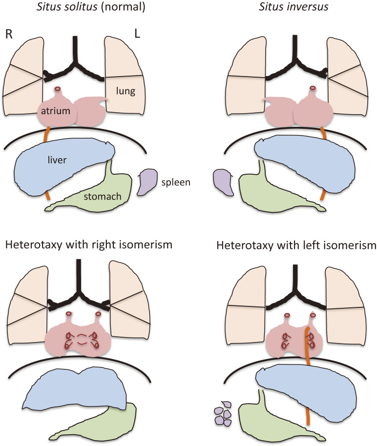
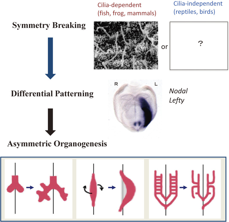
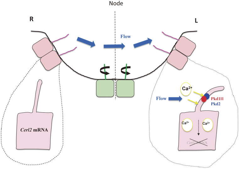
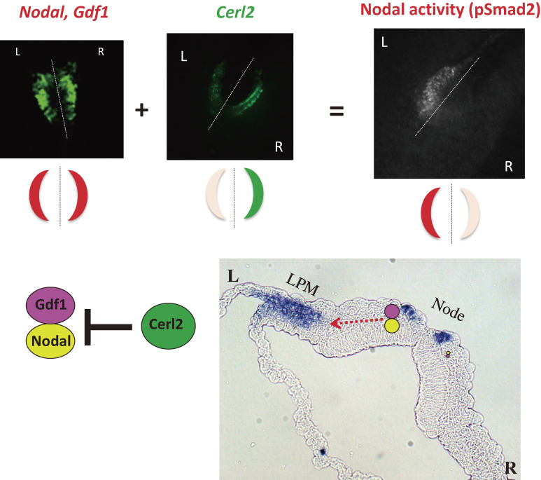
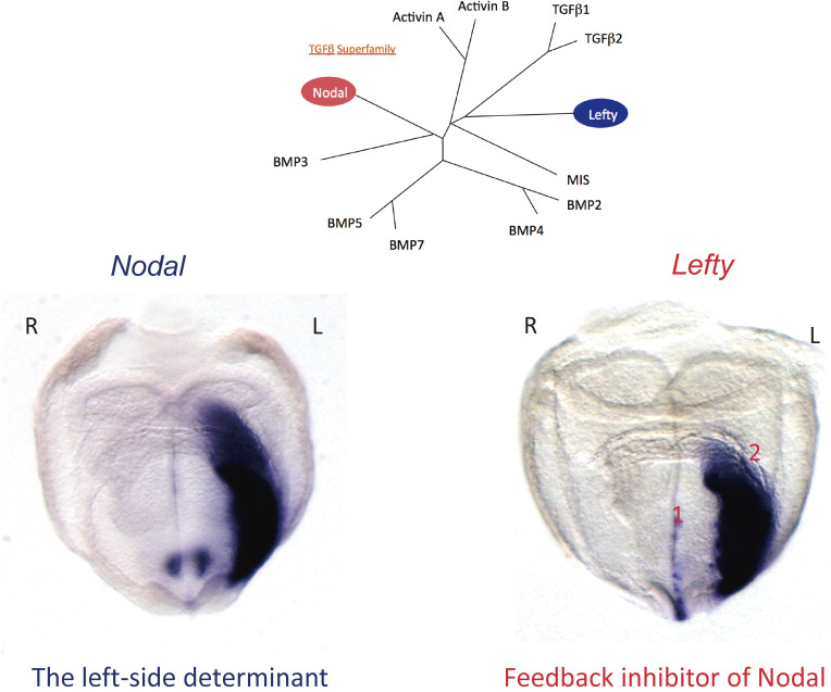
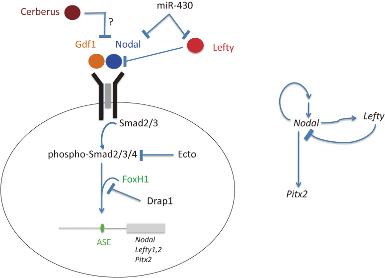
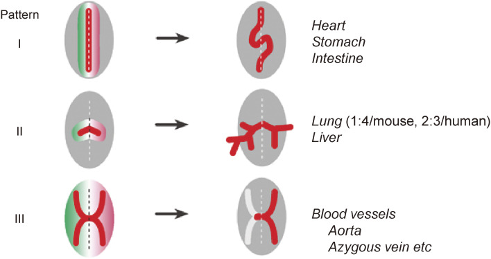
Although the human body appears superficially symmetrical with regard to the left-right (L-R) axis, most visceral organs are asymmetric in terms of their size, shape, or position. Such morphological asymmetries of visceral organs, which are essential for their proper function, are under the control of a genetic pathway that operates in the developing embryo. In many vertebrates including mammals, the breaking of L-R symmetry occurs at a structure known as the L-R organizer (LRO) located at the midline of the developing embryo. This symmetry breaking is followed by transfer of an active form of the signaling molecule Nodal from the LRO to the lateral plate mesoderm (LPM) on the left side, which results in asymmetric expression of Nodal (a left-side determinant) in the left LPM. Finally, L-R asymmetric morphogenesis of visceral organs is induced by Nodal-Pitx2 signaling. This review will describe our current understanding of the mechanisms that underlie the generation of L-R asymmetry in vertebrates, with a focus on mice.

Keywords: Nodal; chirality; cilia; left–right asymmetry.


Figures

Figure 1.
Figure 1.
Schematic illustration of L-R asymmetry of human visceral organs. Normal L-R asymmetry (situs solitus) and three laterality defects that affect the lung, heart, liver, stomach, and spleen are shown. Heterotaxy with right isomerism is usually associated with a bilateral trilobed lung, a large symmetric liver, and the absence of a spleen. Heterotaxy with left isomerism often manifests as a bilateral bilobed lung, multiple spleens, and a pulmonary vein that drains into both the right and left atria.
Figure 2.
Figure 2.
Three steps in the establishment of L-R asymmetry in vertebrates. The three steps include symmetry breaking, patterning, and organogenesis. In the first step, many vertebrates rely on cilia for symmetry breaking, whereas others deploy a cilia-independent, largely unknown mechanism. In the second step, the LPM on the right and left sides undergoes differential patterning as a result of asymmetric Nodal signaling on the left side. In the final step, asymmetric morphogenesis takes place in visceral organs. Modified with permission from Shiratori and Hamada155) and Yoshiba and Hamada.156)
Figure 3.
Figure 3.
Cilia at the node of a mouse embryo as revealed by scanning electron microscopy. a. Lateral view of a mouse embryo at E7.5. The arrow indicates the location of the node. b. Ventral view of the mouse node. The arrow indicates the direction of fluid flow at the node. c. Higher magnification of the node showing the presence of motile cilia. d. Posterior tilt of motile cilia. Clockwise rotation of posteriorly tilted cilia generates fluid flow (leftward) most efficiently when the cilia are farthest from the surface. A, anterior; L, left; P, posterior; R, right. Modified with permission from Shiratori and Hamada.155)
Figure 4.
Figure 4.
Polarization of node cells with motile cilia along the A-P axis. Asymmetric expression of Wnt5 and its inhibitor Sfrp along the A-P axis generates a graded distribution of Wnt5a/b activity (orange). This will induce polarized localization of PCP core proteins (such as Vangl1, 2 in green and Dvl in red) in node cells with motile cilia (pit cells) along the A-P axis of the mouse embryo. Finally, the basal body will be localized to the posterior side of pit cells. Modified with permission from Minegishi et al.42)
Figure 5.
Figure 5.
Immotile cilia at the periphery of the node sense nodal flow. Ciliated cells located in the central region of the node of the mouse embryo (green) possess motile cilia that rotate and generate nodal flow, whereas those located peripherally (pink) possess immotile cilia that sense the flow. Sensing of the flow requires a Pkd2-Pkd1l1 complex with Ca2+ channel activity that is localized to the cilium. The flow-mediated signal, the identity of which remains unknown, triggers the degradation of Cerl2 mRNA preferentially on the left side.
Figure 6.
Figure 6.
Generation of molecular asymmetries at the node. Whereas Nodal mRNA and Gdf1 mRNA are present at similar levels on both sides of the node of a mouse embryo, Cerl2 mRNA shows an asymmetric (R > L) distribution (top panels). Nodal and Gdf1 form a heterodimer that constitutes an active form of Nodal. Given that Cerl2 is an inhibitor of Nodal (bottom left panel), the level of Nodal activity, which is reflected by the abundance of phosphorylated Smad2/3 (pSmad2), shows a R ≪ L pattern (top panels). The Nodal-Gdf1 heterodimer produced by and secreted from perinodal crown cells is thought to be transported to the LPM on the left side via an intraembryonic route (red dotted arrow in the bottom right panel). On reaching the LPM, the Nodal-Gdf1 heterodimer is thought to activate expression of Nodal (indicated by purple staining in the bottom right panel), which is responsive to Nodal signaling. Modified with permission from Shiratori and Hamada,155) Yoshiba and Hamada,156) and Shiratori and Hamada.157)
Figure 7.
Figure 7.
L-R asymmetric expression of Nodal and Lefty in the mouse embryo. Cluster analysis (top) shows the relations among members of the TGFβ superfamily of proteins. In situ hybridization (bottom) reveals the L-R asymmetric expression of Nodal (which encodes a left-side determinant) and Lefty (which encodes a feedback inhibitor of Nodal) in the E8.0 mouse embryo. Note that Nodal and Lefty are expressed in the LPM on the left side, whereas Lefty is also expressed at the midline (left side of the floor plate). There are actually two Lefty genes, with Lefty2 being preferentially expressed in the LPM and Lefty1 at the midline. Modified with permission from Shiratori and Hamada.157)
Figure 8.
Figure 8.
Control of Nodal signaling by various regulators in the developing vertebrate embryo. The Nodal signaling pathway in vertebrate embryos (Nodal → receptor → Smad2/3/4 → FoxH1 → target genes) is regulated in a spatiotemporal manner at multiple levels by various inhibitors including Lefty, Cerberus (Cerl2), Ectodermin (Ecto), Drap1, and miR-430 (left panel). Nodal and Lefty also participate in positive and negative regulatory loops (right panel).
Figure 9.
Figure 9.
Generation of morphological asymmetries. Three different patterns for generation of morphological asymmetries rely on (I) directional looping, (II) differential branching, or (III) one-sided regression. Examples of anatomic structures generated by each mechanism are shown. Modified with permission from Shiratori and Hamada155) and courtesy of Yukio Saijoh (University of Utah).
Figure 10.
Figure 10.
L-R asymmetric expression of Nodal, Lefty, and Pitx2 in the mouse embryo. In situ hybridization reveals that, whereas L-R asymmetric expression of Nodal and Lefty is transient (detectable around E8), L-R asymmetric expression of Pitx2 persists much longer. Asymmetric expression of Nodal and Lefty is controlled by a Nodal-responsive enhancer (ASE), which renders their asymmetric expression transient. Pitx2, however, contains both ASE and an additional enhancer that maintains its expression at later stages. Modified with permission from Shiratori and Hamada.157)
Figure 11.
Figure 11.
L-R asymmetric remodeling of the sixth branchial arch artery in the mouse embryo. In a normal embryo, the right side of the sixth branchial arch artery (BAA) and the right side of the dorsal aorta regress as development proceeds, eventually resulting in arching of the aorta toward the left side. In Pitx2 mutant mice; however, this pattern of remodeling is impaired. AS, aortic sac; RV and LV, right and left ventricle.
Figure 12.
Figure 12.
Rotation and looping of the developing gut. a. Clockwise rotation of the foregut results in translocation of the liver primordium (gray) to the right side of the body cavity. b. Cellular changes that initiate L-R asymmetry in the midgut tube of the chick embryo.120,123) Asymmetries arise in the dorsal mesentery between Hamburger–Hamilton (HH) stages 20 and 22. Left side-specific Nodal-Pitx2 expression drives compaction of mesenchymal cells (green rectangles) within the left dorsal mesentery and promotes retention of a columnar morphology of epithelial cells (blue rectangles) on the left side of the dorsal mesentery. In the right dorsal mesentery, hyaluronan (HA) undergoes modification by Tsg6, which catalyzes the covalent attachment of a heavy chain (orange) of inter-α-trypsin inhibitor. Signals that induce Tsg6 expression in the right half of the dorsal mesentery remain unknown. The modified form of hyaluronan is more stable and accumulates in the right dorsal mesentery, resulting in expansion of mesenchymal cells and exclusion of blood vessels (red line) in the right dorsal mesentery. Together, these cellular asymmetries drive leftward tilting of the gut tube. Left (L) and right (R) sides of the dorsal mesentery are indicated together with the midline (broken line). Modified with permission from Hamada.158)
Figure 13.
Figure 13.
The epithalamic system and L-R asymmetric neural projection in zebrafish. The habenula of zebrafish is divided into a dorsal component and a ventral component (Vhb), with the dorsal habenula being constituted by the lateral (Lhb) and medial (Mhb) subnuclei of unequal size. Further components of the zebrafish epithalamus are the photosensitive pineal (Po) and the asymmetrically organized parapineal organ (Pp). The Lhb and Mhb project to the mesencephalon. The olfactory bulb (Ob) projects to the Lhb and Mhb on the right side. Bilaterally symmetric neural projections are not shown.
Figure 14.
Figure 14.
Mechanisms of L-R asymmetry establishment in various animals. Note that some steps (such as the Nodal-Pitx2 pathway) are conserved, whereas others are divergent.

References

    1. Smith C.A., Sinclair A.H. (2004) Sex determination: Insights from the chicken. Bioessays 26, 120–132. - PubMed
    1. van Soldt B.J., Metscher B.D., Poelmann R.E., Vervust B., Vonk F.J., Müller G.B., et al. (2015) Heterochrony and early left-right asymmetry in the development of the cardiorespiratory system of snakes. PLoS One 10, e116416. - PMC - PubMed
    1. Baillie M. (1789) An account of a remarkable transposition of the viscera in the human body. Lond. Med. J. 10, 178–197. - PMC - PubMed
    1. Kartagener M. (1933) Zur pathogenese der bronchiektasen. 1. Mitteilung: Bronchiektasien bei situs viscerum inversus. Beitr. Klin. Tuberk. 83, 489–501.
    1. Hummel K.P. (1959) Developmental anomalies in mice resulting from action of the gene, disorganization, a semi-dominant lethal. Pediatrics 23, 212–221. - PubMed