

Therapeutic inhibition of microRNA-21 (miR-21) using locked-nucleic acid (LNA)-anti-miR and its effects on the biological behaviors of melanoma cancer cells in preclinical studies
- PMID: 32788885
- PMCID: PMC7418194
- DOI: 10.1186/s12935-020-01394-6
Therapeutic inhibition of microRNA-21 (miR-21) using locked-nucleic acid (LNA)-anti-miR and its effects on the biological behaviors of melanoma cancer cells in preclinical studies
Abstract
Background: Melanoma is a cancer that has a high mortality rate in the absence of targeted therapy. Conventional therapies such as surgery, chemotherapy, and radiotherapy are associated with poor prognosis. The expression of miR-21 appears to be of clinical importance, and the regulation of its expression appears to be an opportunity for treatment.
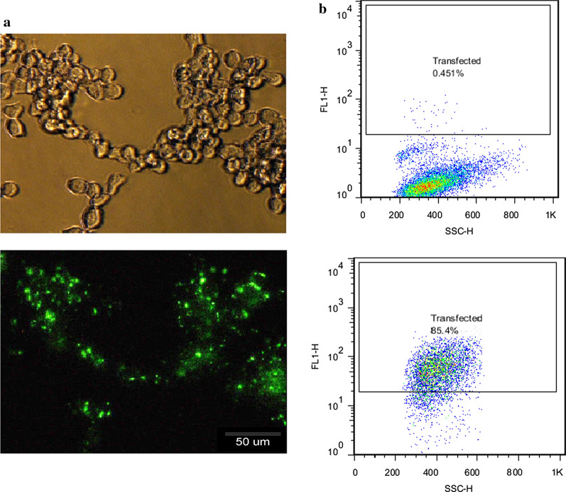
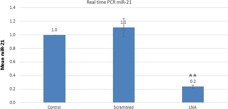
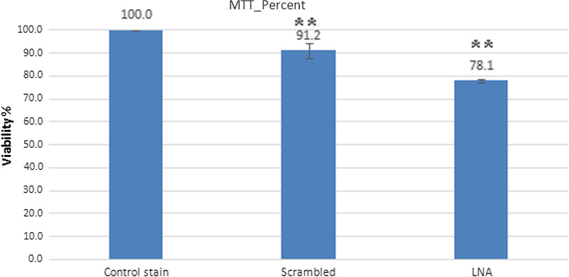
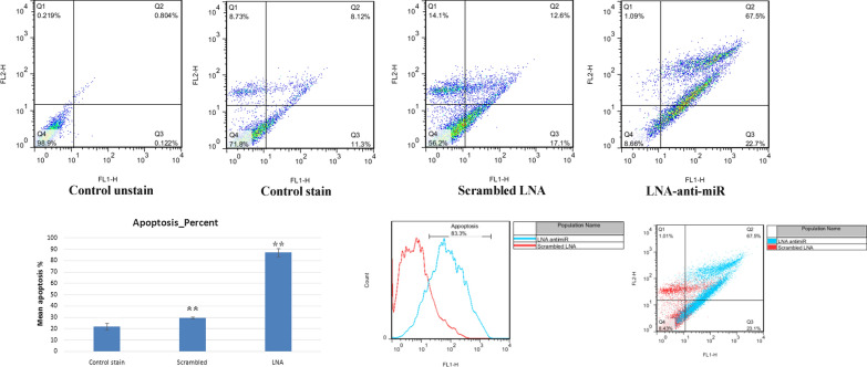
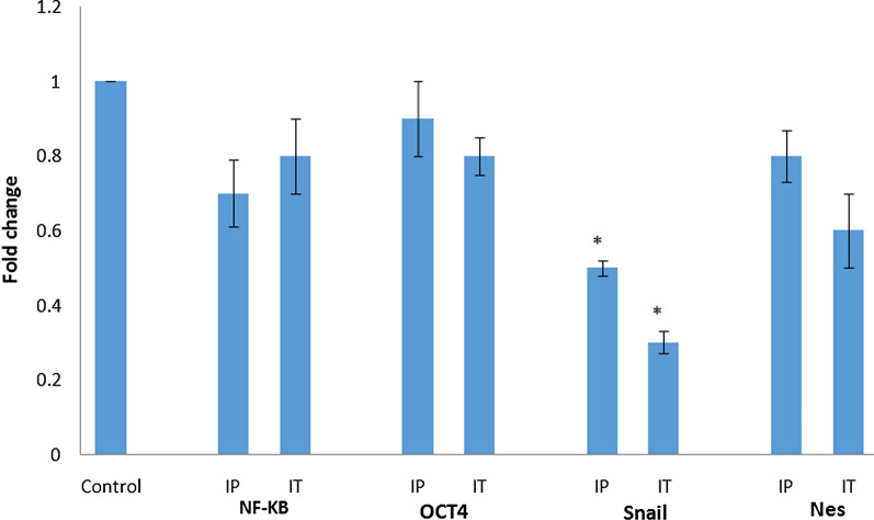
Methods: In this current study, we aimed to evaluate the effects of miR-21 inhibition in- vitro and in-vivo. In-vitro studies have investigated LNA-anti-miR-21 in mouse melanoma cells (B16F10), and in-vivo studies have proposed a model of melanoma in male C57BL/6 mice. To evaluate the anticancer effects of LNA-anti-miR-21, a QRT-PCR analysis was performed using the 2-ΔΔCT method to determine the degree of inhibition of oncomiR-21. The MTT test, propidium iodide/AnnexinV in-vitro, and tumor volume measurement using the QRT-PCR test with the 2-ΔΔCT method were used to estimate the inhibition of miR-21 and the expression of downstream genes including: SNAI1, Nestin (Nes), Oct-4, and NF-kB following miR-21 inhibition. Finally, immunohistochemistry was conducted for an in-vivo animal study.
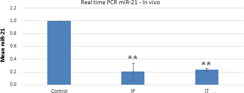
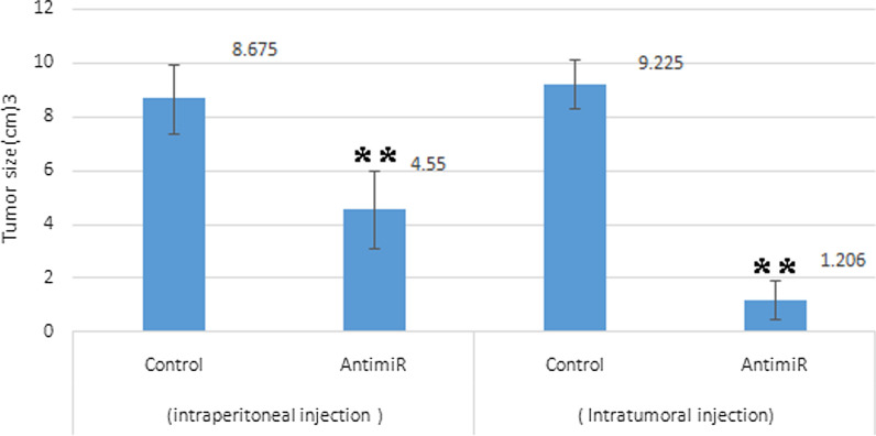
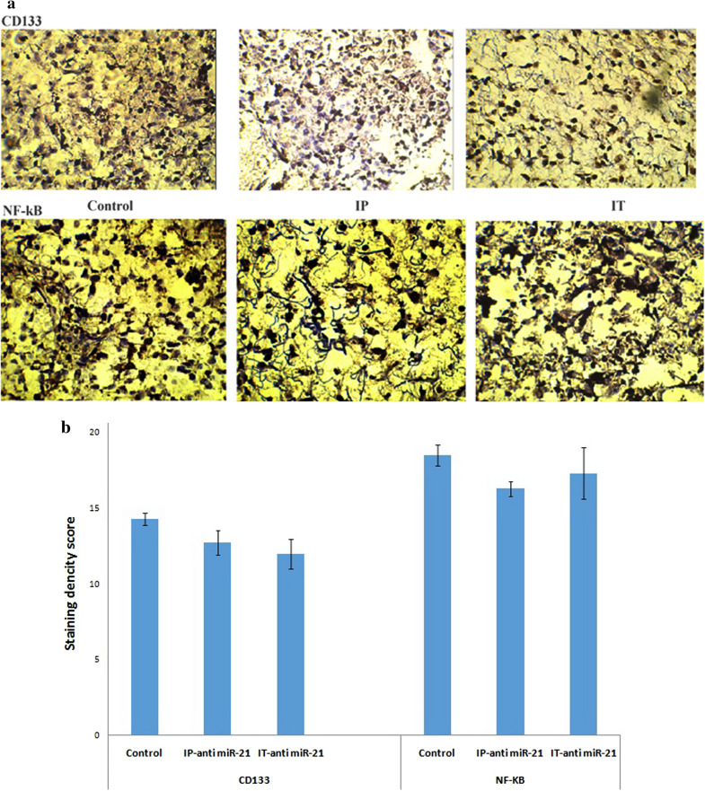
Results: MiR-21 expression was inhibited by 80% after 24 h of B16F10 cell line transfection with LNA-anti-miR-21. The MTT test showed a significant reduction in the number of transfected cells with LNA-anti-miR-21. The transfected cells showed a significant increase in apoptosis in comparison with the control and scrambled LNA groups. According to our in vivo findings, anti-miR-21 could reduce tumor growth and volume in mice receiving intraperitoneal anti-miR after 9 days. The expression of the SNAI1gene was significantly reduced compared to the controls. Immunohistochemical analysis showed no change in CD133 and NF-kB markers.
Conclusion: Our findings suggest LNA-anti-miR-21 can be potentially used as an anticancer agent for the treatment of melanoma.
Keywords: Cutaneous melanoma; LNA-anti-miR-21; miR-21; miRNAs.
© The Author(s) 2020.
Conflict of interest statement
Competing interestsThe authors have declared no conflict of interest.
Figures
References
-
- American Cancer Society. American Cancer Society Cancer Prevention and Early Detection Facts and Figures, Tables & Figures 2020. American Cancer Society; 2020.
-
- Pan B, Lin X, Zhang L, Hong W, Zhang Y. Long noncoding RNA X-inactive specific transcript promotes malignant melanoma progression and oxaliplatin resistance. Melanoma Res. 2019;29(3):254–262. - PubMed
-
- Welsh SJ, Titley J, Brunton L, Valenti M, Monaghan P, Jackman AL, Aherne GW. Comparison of thymidylate synthase (TS) protein up-regulation after exposure to TS inhibitors in normal and tumor cell lines and tissues. Clin Cancer Res. 2000;6(6):2538–2546. - PubMed
LinkOut - more resources
Full Text Sources
Research Materials
