. 2021 Mar 25;23(3):412-421.
doi: 10.1093/neuonc/noaa189.

Functional connectivity within glioblastoma impacts overall survival

Affiliations

Functional connectivity within glioblastoma impacts overall survival

Andy G S Daniel et al. Neuro Oncol. .

Abstract

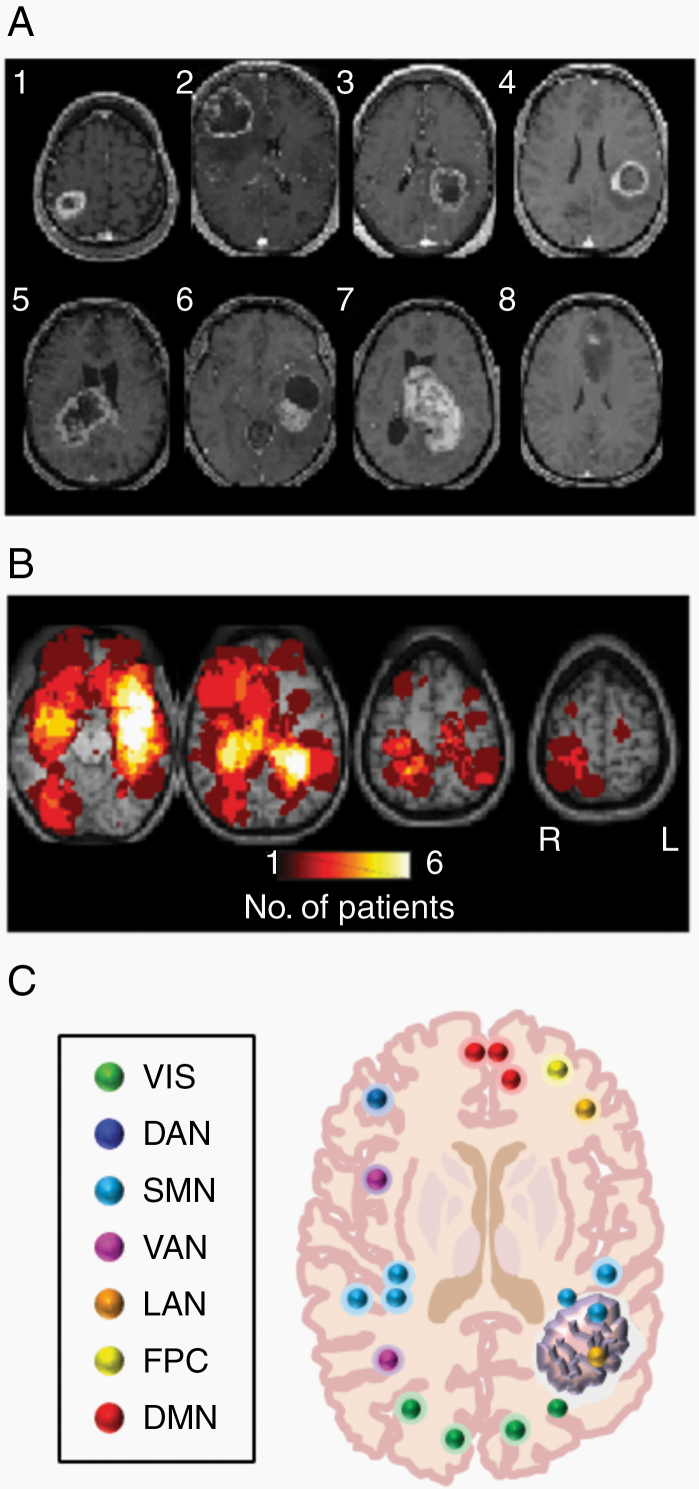
Background: Glioblastoma (GBM; World Health Organization grade IV) assumes a variable appearance on MRI owing to heterogeneous proliferation and infiltration of its cells. As a result, the neurovascular units responsible for functional connectivity (FC) may exist within gross tumor boundaries, albeit with altered magnitude. Therefore, we hypothesize that the strength of FC within GBMs is predictive of overall survival.

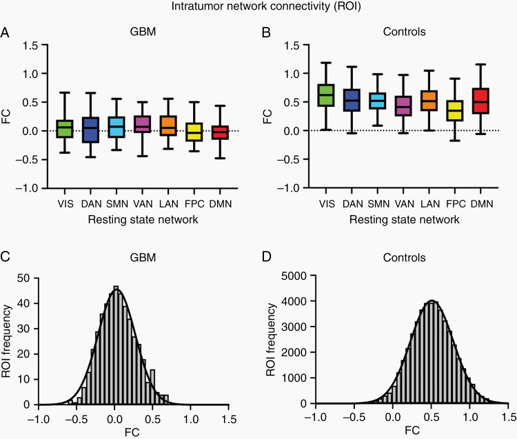
Methods: We used predefined FC regions of interest (ROIs) in de novo GBM patients to characterize the presence of within-tumor FC observable via resting-state functional MRI and its relationship to survival outcomes.

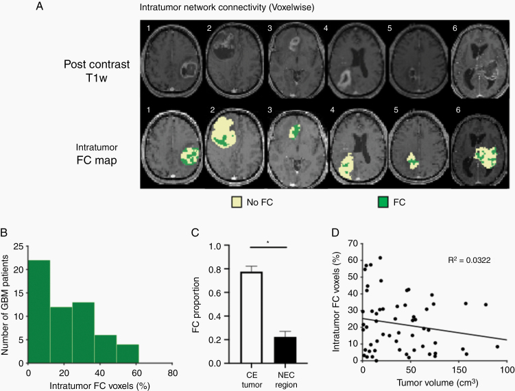
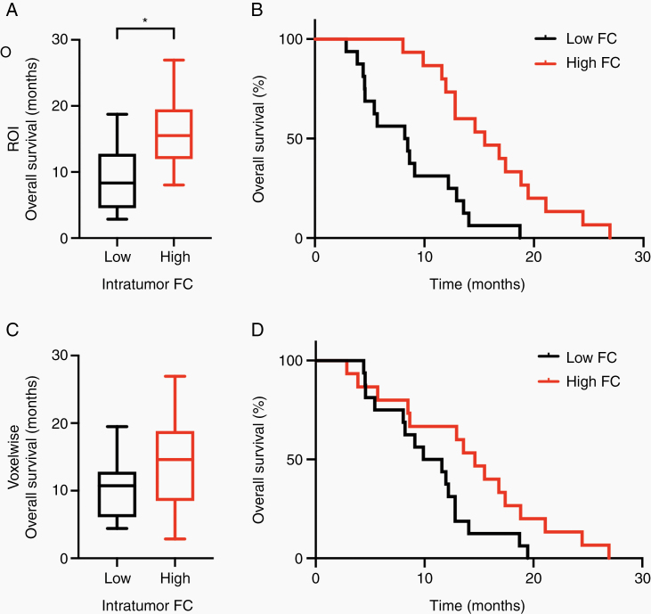
Results: Fifty-seven GBM patients (mean age, 57.8 ± 13.9 y) were analyzed. Functionally connected voxels, not identifiable on conventional structural images, can be routinely found within the tumor mass and was not significantly correlated to tumor size. In patients with known survival times (n = 31), higher intranetwork FC strength within GBM tumors was associated with better overall survival even after accounting for clinical and demographic covariates.

Conclusions: These findings suggest the possibility that functionally intact regions may persist within GBMs and that the extent to which FC is maintained may carry prognostic value and inform treatment planning.

Keywords: FC; functional MRI; glioblastoma; glioma; resting state.


Figures

Fig. 1
Fig. 1
Intratumor FC in GBM patients. (A) Postcontrast T1w images in a sample of 8 patients. (B) Heatmaps showing the distribution of tumor density in the full sample of 57 patients. (C) Schematic of ROIs used to determine network affiliation. Rs-fMRI time series were averaged over ROIs outside the tumor to define RSN-specific time series. Correlation of these time series against intratumor ROIs yielded assessment of intratumor FC. VIS: visual network; DAN: dorsal attention network; SMN: sensorimotor network; VAN: ventral attention network; LAN: language network; FPC: fronto-parietal control network; DMN: default mode network.
Fig. 2
Fig. 2
ROI-based FC (Fisher z-transformed correlation) within GBMs. (A) FC in GBM patients (n = 53). FC strength is represented as a boxplot corresponding to assigned network. The mean of the VIS, DAN, FPN, and DMN FC distributions were not significantly different from zero (one sample t-test, P > 0.05). However, in some patients, some ROIs had FC values of 0.5 or greater. (B) Virtual intratumor FC in controls. Each control (n = 100) was treated as every GBM patient (n = 53) to obtain the expected connectivity strengths and overall intranetwork distributions for the ROIs found within the tumor masks of each GBM patient. The median of every network in controls was greater than its corresponding distribution in GBM patients (two-sample Wilcoxon rank sum-test, P < 0.00001). (C) Intratumor FC distribution in GBM patients, collapsed over RSNs. The mean of this distribution is significantly greater than zero (one-sample t-test, P < 0.0001). (D) Virtual intratumor FC distribution in controls. The mean of this distribution is ~0.5. The control FC distribution is smoother because it represents a larger sample of “intratumor” ROIs.
Fig. 3
Fig. 3
Voxelwise identification of intratumor function. (A) Postcontrast T1w slices and corresponding intratumor FC maps of 6 GBM patients demonstrate tumor and intratumor FC heterogeneity. Green denotes voxels that were assigned FC after obtaining a correlation r > 0.3 with at least one resting-state network. Beige denotes voxels that did not meet this criterion (no FC). (B) Distribution of the percentage of intratumor FC voxels by number of patients reveals that most patients have a low proportion of functional voxels. (C) Bar chart showing the proportion of FC voxels in contrast-enhanced (CE) and necrotic (NEC) areas in GBM tumors (n = 56). Error bars denote 95% CI. CE regions demonstrate significantly higher proportion of FC than NEC areas (CE = 0.78 vs NEC = 0.22, unpaired t-test, P < 0.0001). (D) Plot of percentage of intratumor FC voxels versus tumor volume demonstrating no significant correlation (P = 0.182).
Fig. 4
Fig. 4
Intratumor FC stratifies overall survival in GBM patients. (A) ROI derived FC: Overall survival in GBM patients with low intratumor FC are compared with patients with high intratumor FC. Asterisk indicates significant difference (right-tailed Wilcoxon rank sum, W = 240, P < 0.001, Bonferroni corrected). (B) ROI derived FC: Kaplan–Meier survival analysis comparing overall survival in low intratumor FC GBM patients and high FC patients. Patients with high intratumor FC had a significantly longer overall survival than those with low intratumor FC (HR: 0.25, 95% CI: 0.11–0.58, P = 0.0011). (C) Voxelwise derived FC: Overall survival in GBM patients with low intratumor FC are compared with patients with high intratumor FC (right-tailed Wilcoxon rank sum, W = 281, P = 0.11, Bonferroni corrected). (D) Voxelwise derived FC: Kaplan–Meier survival analysis comparing overall survival in low intratumor FC GBM patients and high FC patients. Patients with high intratumor FC had a significantly longer overall survival than those with low intratumor FC (HR: 0.45, 95% CI: 0.21–0.98, P = 0.044).

References

    1. Wen PY, Kesari S. Malignant gliomas in adults. N Engl J Med. 2008;359(5):492–507. - PubMed
    1. Cuddapah VA, Robel S, Watkins S, Sontheimer H. A neurocentric perspective on glioma invasion. Nat Rev Neurosci. 2014;15(7): 455–465. - PMC - PubMed
    1. Ojemann JG, Miller JW, Silbergeld DL. Preserved function in brain invaded by tumor. Neurosurgery. 1996;39(2):253–25 8; discussion 258. - PubMed
    1. Skirboll SS, Ojemann GA, Berger MS, Lettich E, Winn HR. Functional cortex and subcortical white matter located within gliomas. Neurosurgery. 1996;38(4):678–6 84; discussion 684. - PubMed
    1. Schiffbauer H, Ferrari P, Rowley HA, Berger MS, Roberts TP. Functional activity within brain tumors: a magnetic source imaging study. Neurosurgery. 2001;49(6):1313–13 20; discussion 1320. - PubMed

Publication types