Review
. 2020 Aug 13;4(1):49.
doi: 10.1186/s41747-020-00172-3.

Image-based biomechanical models of the musculoskeletal system

Affiliations
Review

Image-based biomechanical models of the musculoskeletal system

Fabio Galbusera et al. Eur Radiol Exp. .

Abstract

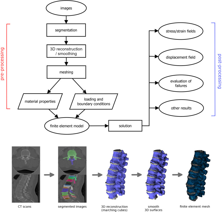
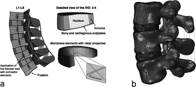
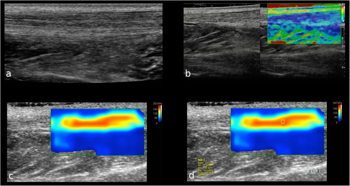
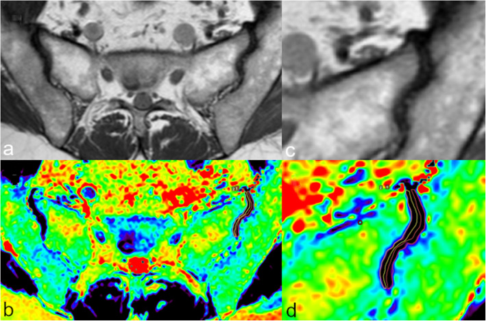
Finite element modeling is a precious tool for the investigation of the biomechanics of the musculoskeletal system. A key element for the development of anatomically accurate, state-of-the art finite element models is medical imaging. Indeed, the workflow for the generation of a finite element model includes steps which require the availability of medical images of the subject of interest: segmentation, which is the assignment of each voxel of the images to a specific material such as bone and cartilage, allowing for a three-dimensional reconstruction of the anatomy; meshing, which is the creation of the computational mesh necessary for the approximation of the equations describing the physics of the problem; assignment of the material properties to the various parts of the model, which can be estimated for example from quantitative computed tomography for the bone tissue and with other techniques (elastography, T1rho, and T2 mapping from magnetic resonance imaging) for soft tissues. This paper presents a brief overview of the techniques used for image segmentation, meshing, and assessing the mechanical properties of biological tissues, with focus on finite element models of the musculoskeletal system. Both consolidated methods and recent advances such as those based on artificial intelligence are described.

Keywords: Artificial intelligence; Finite element analysis; Musculoskeletal System; Tomography; Tomography (x-ray computed), Magnetic resonance imaging.


Conflict of interest statement

The authors declare that they have no competing interests.

Figures

Fig. 1
Fig. 1
The workflow for the development and use of a finite element model from medical images (for example computed tomography scans): segmentation, three-dimensional reconstruction, improvement of the quality of the reconstructed surfaces by means of filtering (smoothing), meshing, assignment of loading/boundary conditions, and of material properties. The model can then be used to make predictions about stresses, strains, displacements, evaluating the failure of the materials, etc. Partially reprinted with permission from [1]
Fig. 2
Fig. 2
Example of segmentation of a knee joint from magnetic resonance imaging scans, manually performed by a human operator, automatically determined by a state-of-the-art deep learning method or with a novel method employing neural networks and a deformable model described in [10]. a Definition of the region of interests for femoral cartilage (green) and tibial cartilage (red); b manual segmentation created by an expert human operator; c automated segmentation obtained with a state-of-the-art deep learning method; d three-dimensional reconstruction of the automated segmentation; e, f filtered images enhancing the contours necessary for the novel approach; g, h segmented slice and three-dimensional reconstruction of the segmentation obtained with the novel method. Reprinted with permission from [10]
Fig. 3
Fig. 3
Comparison of the performance of a tool based on a state-of-the-art deep learning architecture (Deep-Net) [24] and a more conventional, model-based method [25] for the segmentation of computed tomography scans of healthy and pathological spines, highlighting the improved performance of the deep network especially for the pathological cases. Reprinted with permission from [24]
Fig. 4
Fig. 4
Examples of structured mesh on a regular domain (left), structured mesh on an irregular domain (middle) and unstructured mesh on an irregular domain (right). Reprinted with permission from [35]
Fig. 5
Fig. 5
Examples of meshes of a model of the lumbar spine: a structured mesh, highlighting the special techniques used to model the collagen fibre-reinforced nature of the intervertebral disc; b unstructured mesh. a Reprinted with permission from [48]
Fig. 6
Fig. 6
A 31-year-old male patient with healthy left Achilles tendon. a B-mode ultrasound image on longitudinal axis shows the normal thickness and echotexture of the proximal third of the Achilles tendon. b Longitudinal real-time strain sonoelastography shows the normal appearance of the proximal third of the Achilles tendon as blue, which represents stiff tissue. Subcutaneous fat tissue over the tendon appears yellow to green indicating soft tissue. c Shear wave elastography shows that the normal tendon is hard (red) and homogeneous, with the softer tissue over and below the tendon that is easy to distinguish. (d) The box is the region of interest to calculate the tendon elasticity
Fig. 7
Fig. 7
Magnetic resonance imaging of the sacroiliac joints of a 42-year-old female patient. Oblique axial T1-weighted turbo spin-echo image of both sacroiliac joints (a); in detail the left sacroiliac joint (c). The corresponding oblique axial T2 maps (b, d) show the ROIs manually drawn on both sacral and iliac articular side of the joint space of the left sacroiliac joint

References

    1. Galbusera F, Niemeyer F. Chapter 14. Mathematical and finite element modeling. In: Galbusera F, Wilke H-J, editors. Biomechanics of the spine. Cambridge: Academic Press; 2018. pp. 239–255.
    1. Gilbertson LG, Goel VK, Kong WZ, Clausen JD. Finite element methods in spine biomechanics research. Crit Rev Biomed Eng. 1995;23:411–473. doi: 10.1615/critrevbiomedeng.v23.i5-6.20. - DOI - PubMed
    1. Zienkiewicz OC, Taylor RL, Nithiarasu P, Zhu JZ. The finite element method. London: McGraw-Hill; 1977.
    1. Reddy JN. An introduction to the finite element method. New York: McGraw-Hill Education; 1993.
    1. Lalitha M, Kiruthiga M, Loganathan C. A survey on image segmentation through clustering algorithm. Int J Sci Res. 2013;2:348–358.

Publication types

MeSH terms