. 2020 Aug 13;16(8):e1008760.
doi: 10.1371/journal.ppat.1008760. eCollection 2020 Aug.

Cell type- and replication stage-specific influenza virus responses in vivo

Affiliations

Cell type- and replication stage-specific influenza virus responses in vivo

Elizabeth J Fay et al. PLoS Pathog. .

Abstract

Influenza A viruses (IAVs) remain a significant global health burden. Activation of the innate immune response is important for controlling early virus replication and spread. It is unclear how early IAV replication events contribute to immune detection. Additionally, while many cell types in the lung can be infected, it is not known if all cell types contribute equally to establish the antiviral state in the host. Here, we use single-cycle influenza A viruses (scIAVs) to characterize the early immune response to IAV in vitro and in vivo. We found that the magnitude of virus replication contributes to antiviral gene expression within infected cells prior to the induction of a global response. We also developed a scIAV that is only capable of undergoing primary transcription, the earliest stage of virus replication. Using this tool, we uncovered replication stage-specific responses in vitro and in vivo. Using several innate immune receptor knockout cell lines, we identify RIG-I as the predominant antiviral detector of primary virus transcription and amplified replication in vitro. Through a Cre-inducible reporter mouse, we used scIAVs expressing Cre-recombinase to characterize cell type-specific responses in vivo. Individual cell types upregulate unique sets of antiviral genes in response to both primary virus transcription and amplified replication. We also identified antiviral genes that are only upregulated in response to direct infection. Altogether, these data offer insight into the early mechanisms of antiviral gene activation during influenza A infection.


Conflict of interest statement

The authors have declared that no competing interests exist.

Figures
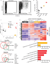
Fig 1
Fig 1. Heterogeneous antiviral response to scIAV infection at 12 hpi.
B6 mice were infected with 105 PFU of scIAV-ΔHA-mCherry or 103 PFU PR8. (A) CD45-CD31- cells were analyzed for mCherry expression at 12 hpi. Data representative of six independent experiments with n = 3–4 mice per group. mCherry negative, low, and high CD45-CD31-cells were sorted at 12 hpi for RNA-seq analysis. (B) IAV CPM (C) MDS of naïve and mCherry negative, low, and high cells. (D) Heatmap of 207 ISGs differentially expressed in the indicated populations. Cutoff of false discovery rate (FDR) is ≤ 0.05. (E) Overlapping low ISGs (mCherry 24h cluster 2, GFP cluster 1, and mCherry 12h clusters 1 and 5) and high ISGs (mCherry 24h cluster 4, GFP cluster 4b, and mCherry 12h cluster 8). Only genes induced to ≥10 CPM in at least one sample are shown. (F) Gene ontology analysis (DAVID, biological processes) was performed for genes upregulated in mCherry high, low, and negative cells over naïve (logFC ≥ 1.5, FDR ≤ 0.05). Unique pathways identified in mCherry low (top) mCherry high (bottom) are shown (FDR ≤ 0.05). (B-F) representative of one experiment with n = 3 mice per group.
Fig 2
Fig 2. Genetic restriction of IAV to primary transcription.
(A) A549 cells were infected with ΔHA-mCherry or ΔPB1-mCherry at MOI = 1 and harvested at the indicated timepoints for RNA-seq. Positive sense (m/cRNA) and negative sense (vRNA) RNA was quantified. Data representative of two independent experiments with n = 3 replicate samples per group. (B) vRNA was extracted from the indicated virus stock and PB2 (top) and NA (bottom) were amplified. Representative of two independent experiments. (C) 1.24x10⁵ PFU of ΔPB1-Cre and ΔHA-Cre virus were analyzed for PB1 protein. Representative of three independent experiments.
Fig 3
Fig 3. Primary transcription is detected by RIG-I in vitro.
Indicated A549 cells were infected with ΔHA-mCherry or ΔPB1-mCherry at MOI = 1 and RNA extracted at 12 hpi for mRNA-seq analysis. (A) Total (black) and ISGs (red) differentially expressed genes are shown. The number of genes significantly upregulated (logFC≥2, FDR≤0.05, log CPM≥1.5) over naïve is shown in upper right of plot. The highest upregulated genes (logFC≥5, logCPM≥5) are listed in each plot. (B) Normalized expression level (read CPM) for IFNB1 and IFNL3 in naïve and ΔHA-mCherry or ΔPB1-mCherry infected A549 cells. Data representative of one experiment with n = 3 replicate samples per group.
Fig 4
Fig 4. Detection of cells supporting only primary transcription in vivo.
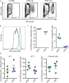
(A-D) Cre reporter mice were infected with 103 PFU of PR8 or 105 PFU of ΔHA-Cre or ΔPB1-Cre and lungs analyzed by flow cytometry at 24 hpi. (A-B) Epithelial cells (CD45-CD31-) were analyzed for tdTomato expression. (C) Number and percentage of CD45-CD31- epithelial cells that are tdTomato+. (D) The percentage of infected (tdTomato+) ATI, ATII, and ATII cells was quantified. (A-D) representative of 3 independent experiments with n = 3–4 mice.
Fig 5
Fig 5. Transcriptional response to viral replication and primary transcription in epithelial cell subsets in vivo.
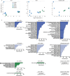
Cre reporter mice were infected with 105 pfu ΔHA-Cre or ΔPB1-Cre and tdTomato+ and tdTomato- ATI, ATII, and ciliated cells were sorted at 24 hpi for RNAseq analysis. (A) MDS plots of the indicated cell type. ΔHA+: infected, ΔHA-: uninfected, ΔPB1+: infected, ΔPB1-: uninfected. (B) GO analysis (DAVID, biological processes) of significantly upregulated genes (logFC≥1.5, FDR≤0.01, CPM≥10 in at least one sample) for the indicated population of each cell type. Significantly enriched pathways (FDR≤0.05) are shown.
Fig 6
Fig 6. Cell type-specific antiviral responses to stages of virus replication in vivo.
RNA-seq data from Fig 5 were analyzed for ISG expression (A) Unique and overlapping ISGs significantly upregulated (logFC≥1.5, FDR ≤0.05, CPM≥10 in at least one population) in ΔHA- or ΔPB1-infected tdTomato+ cells compared to naïve indicated cell type. (B) Unique and overlapping ISGs significantly upregulated in ΔHA-infected tdTomato+ cells. (C) Infection-specific ISGs were identified by comparing genes in tdTomato+ (ΔHA/ΔPB1+) and tdTomato- (ΔHA/PB1-) cells. Select genes in each population listed. Data representative of one independent experiment with n = 2–6 mice per group.

References

    1. Benitez AA, Panis M, Xue J, Varble A, Shim JV, Frick AL, et al. In Vivo RNAi Screening Identifies MDA5 as a Significant Contributor to the Cellular Defense against Influenza A Virus. Cell Rep. 2015;11(11):1714–26. 10.1016/j.celrep.2015.05.032 - DOI - PMC - PubMed
    1. Loo YM, Fornek J, Crochet N, Bajwa G, Perwitasari O, Martinez-Sobrido L, et al. Distinct RIG-I and MDA5 signaling by RNA viruses in innate immunity. Journal of virology. 2008;82(1):335–45. 10.1128/JVI.01080-07 - DOI - PMC - PubMed
    1. Te Velthuis AJ, Fodor E. Influenza virus RNA polymerase: insights into the mechanisms of viral RNA synthesis. Nature reviews Microbiology. 2016;14(8):479–93. 10.1038/nrmicro.2016.87 - DOI - PMC - PubMed
    1. York A, Hengrung N, Vreede FT, Huiskonen JT, Fodor E. Isolation and characterization of the positive-sense replicative intermediate of a negative-strand RNA virus. Proceedings of the National Academy of Sciences of the United States of America. 2013;110(45):E4238–45. 10.1073/pnas.1315068110 - DOI - PMC - PubMed
    1. Vreede FT, Jung TE, Brownlee GG. Model suggesting that replication of influenza virus is regulated by stabilization of replicative intermediates. Journal of virology. 2004;78(17):9568–72. 10.1128/JVI.78.17.9568-9572.2004 - DOI - PMC - PubMed

Publication types

MeSH terms