. 2020 Aug 13;15(8):e0237514.
doi: 10.1371/journal.pone.0237514. eCollection 2020.

Human Golgi phosphoprotein 3 is an effector of RAB1A and RAB1B

Affiliations

Human Golgi phosphoprotein 3 is an effector of RAB1A and RAB1B

Viviana A Cavieres et al. PLoS One. .

Abstract

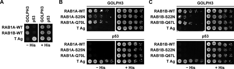
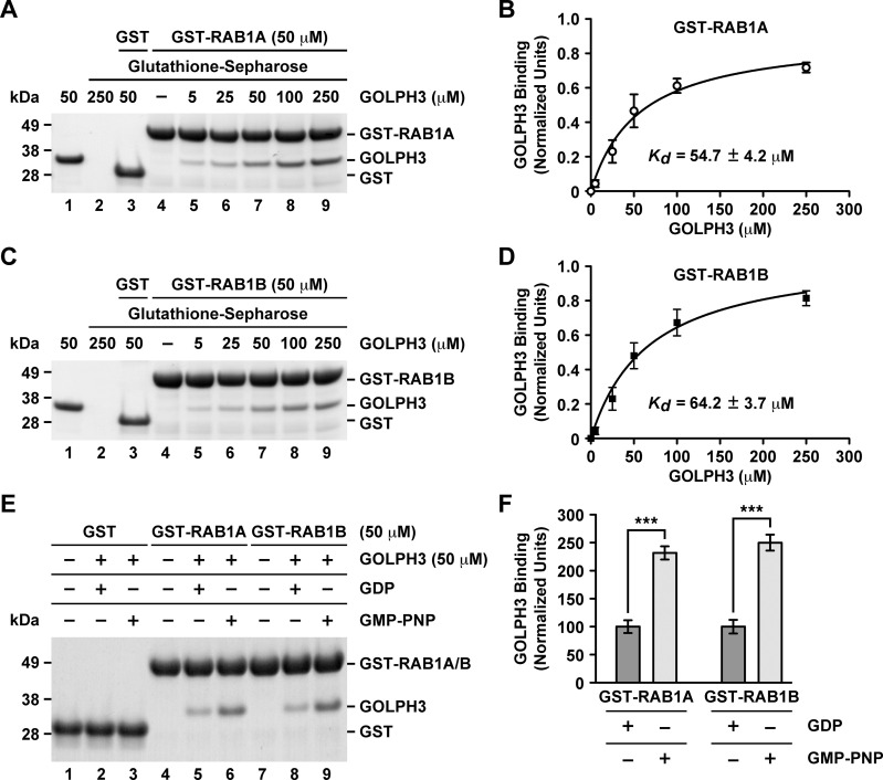
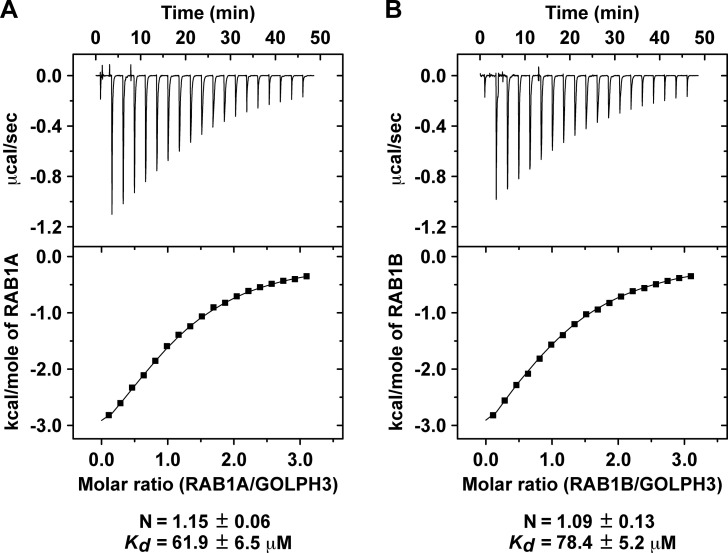
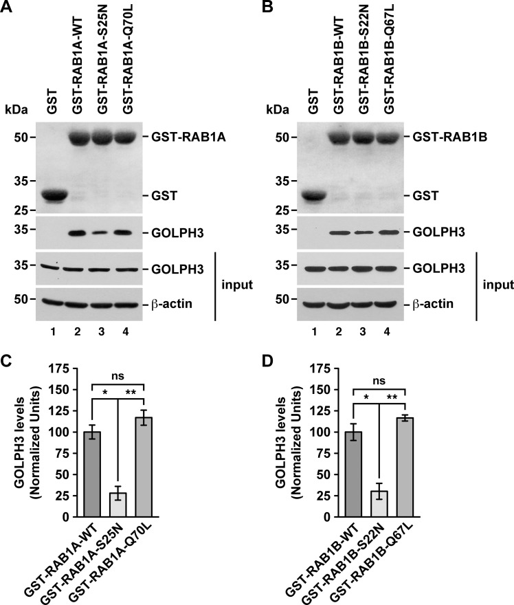
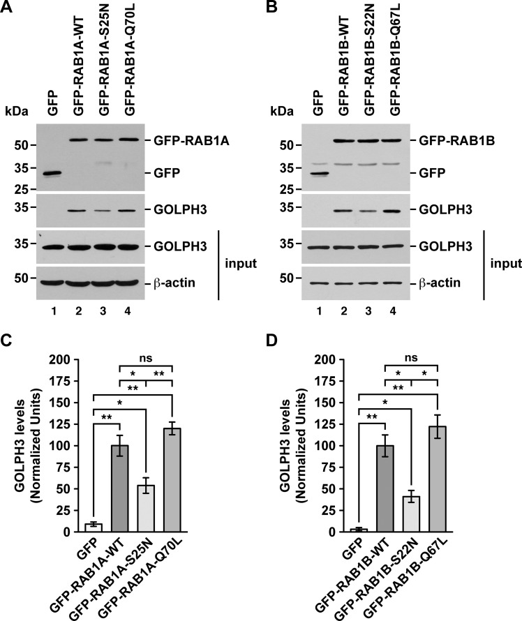
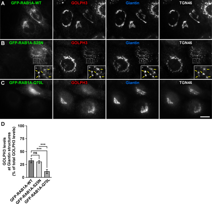
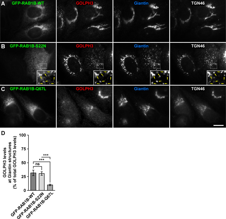
Golgi phosphoprotein 3 (GOLPH3) is a peripheral membrane protein localized at the trans-Golgi network that is also distributed in a large cytosolic pool. GOLPH3 has been involved in several post-Golgi protein trafficking events, but its precise function at the molecular level is not well understood. GOLPH3 is also considered the first oncoprotein of the Golgi apparatus, with important roles in several types of cancer. Yet, it is unknown how GOLPH3 is regulated to achieve its contribution in the mechanisms that lead to tumorigenesis. Binding of GOLPH3 to Golgi membranes depends on its interaction to phosphatidylinositol-4-phosphate. However, an early finding showed that GTP promotes the binding of GOLPH3 to Golgi membranes and vesicles. Nevertheless, it remains largely unknown whether this response is consequence of the function of GTP-dependent regulatory factors, such as proteins of the RAB family of small GTPases. Interestingly, in Drosophila melanogaster the ortholog of GOLPH3 interacts with- and behaves as effector of the ortholog of RAB1. However, there is no experimental evidence implicating GOLPH3 as a possible RAB1 effector in mammalian cells. Here, we show that human GOLPH3 interacted directly with either RAB1A or RAB1B, the two isoforms of RAB1 in humans. The interaction was nucleotide dependent and it was favored with GTP-locked active state variants of these GTPases, indicating that human GOLPH3 is a bona fide effector of RAB1A and RAB1B. Moreover, the expression in cultured cells of the GTP-locked variants resulted in less distribution of GOLPH3 in the Golgi apparatus, suggesting an intriguing model of GOLPH3 regulation.


Conflict of interest statement

The authors have declared that no competing interests exist.

Figures

Fig 1
Fig 1. Yeast two-hybrid analysis of the interaction of GOLPH3 with either RAB1A or RAB1B.
(A-C) Yeast were co-transformed with plasmids encoding either Gal4bd fused to human GOLPH3 or Gal4ad fused to either RAB1A or RAB1B wild type variants (RAB1A-WT and RAB1B-WT; A), or to the GDP-locked inactive state variants of either RAB1A or RAB1B (RAB1A-S25N and RAB1B-S22N; B and C), or to the respective GTP-locked active state variants (RAB1A-Q70L and RAB1B-Q67L; B and C). Mouse p53 fused to Gal4bd and SV40 large T antigen (T Ag) fused to Gal4ad were used as controls. Co-transformed cells were spotted onto His-deficient (-His) or His-containing (+His) plates and incubated at 30°C. Growth is indicative of interactions. The figure depicts representative images of three independent experiments.
Fig 2
Fig 2. GST-pulldown analysis of the interaction of GOLPH3 with either RAB1A or RAB1B.
(A and C) GST-pulldown (A and C, lanes 2–9) using GST-RAB1A at 50 μM (A, lanes 4–9) or GST-RAB1B at 50 μM (C, lanes 4–9) with the indicated different concentrations of GOLPH3 (A and C, lanes 2, 3, 5–9). Lane 1 on each panel was loaded with the indicated amount of GOLPH3 that is equivalent to the amount that was used for the pulldown loaded in lane 7 on each panel. Lanes 2–4 are control pulldowns: lanes 2 are pulldowns using the indicated concentration of GOLPH3 in the absence either of GST-RAB1A (A) or of GST-RAB1B (C); lanes 3 are pulldowns using GST and the indicated concentration of GOLPH3; and lanes 4 are pulldowns using the indicated concentration either of GST-RAB1A (A) or of GST-RAB1B (C) in the absence of GOLPH3. Proteins on glutathione-Sepharose beads were eluted with sample buffer, separated by SDS-PAGE, and stained with Coomassie Blue G-250. The position of the proteins used is indicated on the right. The position of molecular mass markers is indicated on the left. (B and D) Densitometry quantification of the amount of GOLPH3 pulled down as shown in A and C. The amount of GOLPH3 was normalized to the amount in each lane either of GST-RAB1A (B) or of GST-RAB1B (D), and plotted versus each GOLPH3 concentration (n = 3 independent experiments). The data was fitted assuming a 1:1 stoichiometry resulting in the indicated KD. (E) GST-pulldown of GOLPH3 after GDP or GMP-PNP nucleotide exchange on GST-RAB1A (lanes 4–6) or on GST-RAB1B (lanes 7–9). Mock nucleotide exchanges on GST were used as controls (lanes 1–3). (F) Densitometry quantification of the amount of GOLPH3 pulled down as shown in E. The amount of GOLPH3 was normalized to the amount in each lane either of GST-RAB1A (lanes 5 and 6; n = 3 independent experiments) or of GST-RAB1B (lanes 8 and 9; n = 3 independent experiments).
Fig 3
Fig 3. Isothermal titration calorimetry analysis of the interaction of GOLPH3 with either RAB1A or RAB1B.
(A-B) Isothermal titration calorimetry of GOLPH3 either with RAB1A (A) or with RAB1B (B). The stoichiometry (N) and KD for each interaction are expressed as the mean ± SEM (n = 3).
Fig 4
Fig 4. GST-pulldown analysis of the interaction of endogenous GOLPH3 with either RAB1A or RAB1B.
(A-B) Pulldowns of the indicated variants of GST-RAB1A (A) or of GST-RAB1B (B) incubated with soluble protein extracts from human neuroglioma H4 cells. Samples of the pulldowns (upper two panels) and of the protein extracts used in the pulldowns (lower two panels; input) were processed by SDS-PAGE followed by immunoblotting. Before the immunoblotting of the pulled down samples, nitrocellulose membranes were subjected to Ponceau S staining (upper panels). The position of GST (used as pulldown control) and of the GST-tagged RAB1A or RAB1B variants is indicated on the right. Immunoblottings were carried out using antibodies to detect the proteins indicated on the right of the bottom three panels. The position of molecular mass markers is indicated on the left. (C-D) Densitometry quantification of the amount of GOLPH3 pulled down as shown in the second panel of A and B. The immunoblot signal of anti-β-actin was used as loading control. Bar represents the mean ± standard deviation (n = 3 independent experiments). * P < 0.05; ** P < 0.01; ns, not statistically significant.
Fig 5
Fig 5. GFP-Trap analysis of the interaction of endogenous GOLPH3 with either RAB1A or RAB1B.
(A-B) Soluble protein extracts from human H4 neuroglioma cells transiently expressing the indicated variants of GFP-RAB1A (A) or of GFP-RAB1B (B) were subjected to the GFP-Trap assay. Samples of the GFP traps (upper two panels) and of the protein extracts used in the pulldowns (lower two panels; input) were processed by SDS-PAGE followed by immunoblotting. Immunoblottings were carried out using antibodies to detect the proteins indicated on the right. Antibody to GFP was used to detect GFP (used as GFP-Trap control) and the GFP-tagged RAB1A and RAB1B variants. The position of molecular mass markers is indicated on the left. (C-D) Densitometry quantification of the amount of GOLPH3 pulled down as shown in the third panel of A and B. The immunoblot signal of anti-β-actin was used as loading control. Bar represents the mean ± standard deviation (n = 3 independent experiments). * P < 0.05; ** P < 0.01; ns, not statistically significant.
Fig 6
Fig 6. Fluorescence microscopy analysis of the effect of the expression of GFP-tagged RAB1A variants on GOLPH3 subcellular distribution.
(A-C) H4 cells grown in glass coverslips were transfected to express either of the indicated GFP-tagged variant of RAB1A (green channels). Cells were fixed, permeabilized, and triple-labeled with rabbit polyclonal antibody to GOLPH3, mouse monoclonal antibody to Giantin and sheep polyclonal antibody to TGN46. Secondary antibodies were Alexa-Fluor-594-conjugated donkey anti-rabbit IgG (red channels), Alexa-Fluor-647-conjugated donkey anti-mouse IgG (blue channels) and Alexa-Fluor-350-conjugated donkey anti-sheep IgG (white channels). Stained cells were examined by fluorescence microscopy. Insets in B: X3 magnification, with arrows indicating colocalization at Golgi punctae. Bar, 10 μm. (D) Quantification as described in Materials and Methods of the percentage of fluorescence signal of anti-GOLPH3 associated to Golgi elements decorated with anti-Giantin. Bar represents the mean ± standard deviation (n = 3 independent experiments, and 15 cells in each experiment were analyzed). *** P < 0.001; ns, not statistically significant.
Fig 7
Fig 7. Fluorescence microscopy analysis of the effect of the expression of GFP-tagged RAB1B variants on GOLPH3 subcellular distribution.
(A-C) H4 cells grown in glass coverslips were transfected to express either of the indicated GFP-tagged variant of RAB1B (green channels). Cells were fixed, permeabilized, and triple-labeled with rabbit polyclonal antibody to GOLPH3, mouse monoclonal antibody to Giantin and sheep polyclonal antibody to TGN46. Secondary antibodies were Alexa-Fluor-594-conjugated donkey anti-rabbit IgG (red channels), Alexa-Fluor-647-conjugated donkey anti-mouse IgG (blue channels) and Alexa-Fluor-350-conjugated donkey anti-sheep IgG (white channels). Stained cells were examined by fluorescence microscopy. Insets in B: X3 magnification, with arrows indicating colocalization at Golgi punctae. Bar, 10 μm. (D) Quantification as described in Materials and Methods of the percentage of fluorescence signal of anti-GOLPH3 associated to Golgi elements decorated with anti-Giantin. Bar represents the mean ± standard deviation (n = 3 independent experiments, and 15 cells in each experiment were analyzed). *** P < 0.001; ns, not statistically significant.

References

    1. Lanzetti L, Di Fiore PP. Behind the Scenes: Endo/Exocytosis in the Acquisition of Metastatic Traits. Cancer research. 2017;77(8):1813–7. 10.1158/0008-5472.CAN-16-3403 . - DOI - PubMed
    1. Wu CC, Taylor RS, Lane DR, Ladinsky MS, Weisz JA, Howell KE. GMx33: a novel family of trans-Golgi proteins identified by proteomics. Traffic. 2000;1(12):963–75. . - PubMed
    1. Bell AW, Ward MA, Blackstock WP, Freeman HN, Choudhary JS, Lewis AP, et al. Proteomics characterization of abundant Golgi membrane proteins. The Journal of biological chemistry. 2001;276(7):5152–65. 10.1074/jbc.M006143200 . - DOI - PubMed
    1. Snyder CM, Mardones GA, Ladinsky MS, Howell KE. GMx33 associates with the trans-Golgi matrix in a dynamic manner and sorts within tubules exiting the Golgi. Molecular biology of the cell. 2006;17(1):511–24. 10.1091/mbc.e05-07-0682 - DOI - PMC - PubMed
    1. Dippold HC, Ng MM, Farber-Katz SE, Lee SK, Kerr ML, Peterman MC, et al. GOLPH3 bridges phosphatidylinositol-4- phosphate and actomyosin to stretch and shape the Golgi to promote budding. Cell. 2009;139(2):337–51. 10.1016/j.cell.2009.07.052 - DOI - PMC - PubMed

Publication types