. 2020 Aug 3;61(10):27.
doi: 10.1167/iovs.61.10.27.

Epithelial Cell Migration and Proliferation Patterns During Initial Wound Closure in Normal Mice and an Experimental Model of Limbal Stem Cell Deficiency

Affiliations

Epithelial Cell Migration and Proliferation Patterns During Initial Wound Closure in Normal Mice and an Experimental Model of Limbal Stem Cell Deficiency

Sudan Puri et al. Invest Ophthalmol Vis Sci. .

Abstract

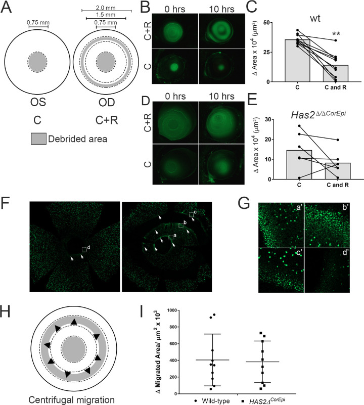
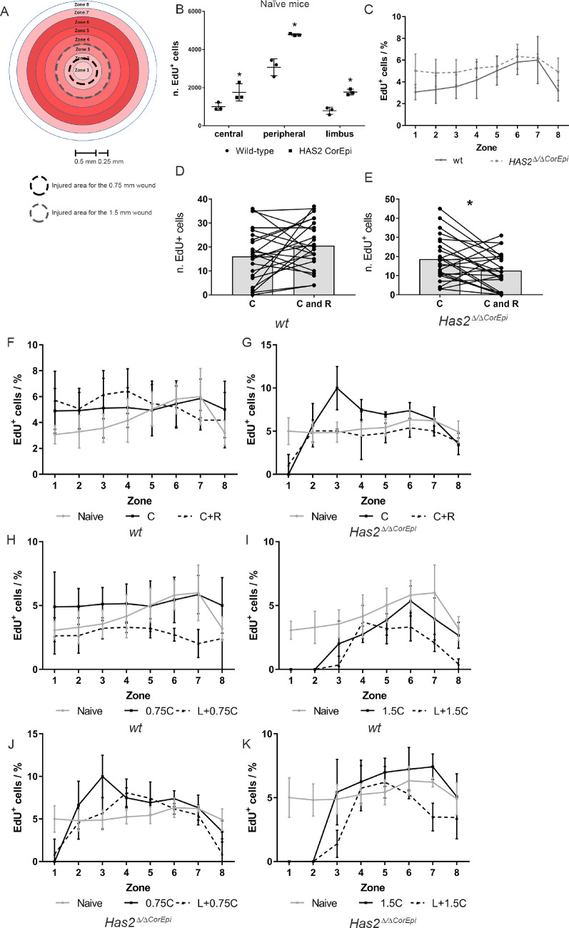
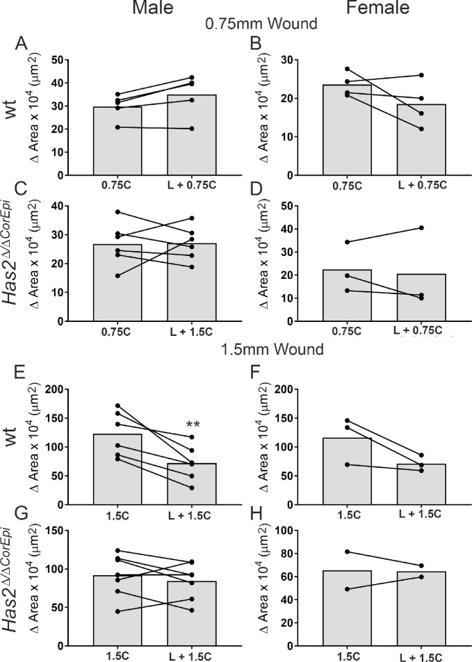
Purpose: Establishing the dynamics of corneal wound healing is of vital importance to better understand corneal inflammation, pathology, and corneal regeneration. Numerous studies have made great strides in investigating multiple aspects of corneal wound healing; however, some aspects remain to be elucidated. This study worked toward establishing (1) if epithelial limbal stem cells (LSCs) are necessary for healing all corneal wounds, (2) the mechanism by which epithelial cells migrate toward the wound, and (3) if centrifugal epithelial cell movement exists.

Methods: To establish different aspects of corneal epithelial wound healing we subjected mice lacking hyaluronan synthase 2 (previously shown to lack LSCs) and wild-type mice to different corneal debridement injury models.

Results: Our data show that both LSCs and corneal epithelial cells contribute toward closure of corneal wounds. In wild-type mice, removal of the limbal rim delayed closure of 1.5-mm wounds, and not of 0.75-mm wounds, indicating that smaller wounds do not rely on LSCs as do larger wounds. In mice shown to lack LSCs, removal of the limbal rim did not affect wound healing, irrespective of the wound size. Finally, transient amplifying cells and central epithelial cells move toward a central corneal wound in a centripetal manner, whereas central epithelial cells may move in a centrifugal manner to resurface peripheral corneal wounds.

Conclusions: Our findings show the dimensions of the corneal wound dictate involvement of LSCs. Our data suggest that divergent findings by different groups on the dynamics of wound healing can be in part owing to differences in the wounding models used.


Conflict of interest statement

Disclosure: S. Puri, None; M. Sun, None; K.N. Mutoji, None; T.F. Gesteira, Co-Founder of and is employed by Optimvia LCC; V.J. Coulson-Thomas, None

Figures

Figure 1.
Figure 1.
The effect of a peripheral ring wound on healing a central corneal wound in wild-type and Has2Δ/ΔCorEpi mice. (A) Schematic of the wounding model; the left eye was subjected to a central circular wound of 0.75 mm in diameter, while the right eye was subjected to the same central wound, as well as a ring wound in the peripheral cornea (C+R). The debrided area is represented in gray and spared epithelium in white. (B) Images were acquired of the ocular surface of wild-type mice treated with fluorescein under a stereomicroscope using the GFP filter immediately and 10 hours after injury. (C) The wounded area of wild-type mice was measured immediately and 10 hours after injury and the healed area calculated. Central wounds surrounded by a peripheral ring wound (C+R) presented reduced healed area when compared with corneas with only a circular wound (C). (D) Images were acquired of the ocular surface of Has2Δ/ΔCorEpi mice treated with fluorescein under a stereomicroscope using the GFP filter immediately and 10 hours after injury. (E) The wounded area was measured immediately and 10 hours after injury and the healed area calculated. The ring wound surrounding the central circular wound (C+R) did not significantly delay wound healing. (F) Images were acquired of EdU (green) stained whole mounted corneas, demarcated areas are shown in higher magnification in (G), corneas were counter stained with DAPI. (H) Schematic representing the centrifugal movement of epithelial cells that was assayed. (I) The migrated area of the wound edge of the innermost rim of the ring wound was calculated for wild-type and Has2Δ/ΔCorEpi mice to verify whether the ring wound also healed in the central to limbal direction, indicating epithelial cells within the central area are capable of moving in a centrifugal manner. Both wild-type and Has2Δ/ΔCorEpi mice presented centrifugal epithelial cell movement. No statistically significant difference was found in the centrifugal migrated area between wild-type and Has2Δ/ΔCorEpi mice. Fluorescein was used to visualize the wounded area and also for the investigator to ascertain the successful removal of cells within the area demarcated with a trephine. Because fluorescein may diffuse throughout the cornea while verifying the quality of the injury, the injured area was measured from the wound edges. *P ≤ 0.05 and **P ≤ 0.01.
Figure 2.
Figure 2.
Comparison of the healed area of central wounds surrounded or not surrounded by peripheral ring wounds in Has2Δ/ΔCorEpi and wild-type mice by gender. The healed area was compared for central wounds surrounded or not surrounded by a peripheral ring wound (C and C+R, respectively) with gender as a variable. (A) Male wild-type mice, (B) female wild-type mice, (C) male Has2Δ/ΔCorEpi mice, and (D) female Has2Δ/ΔCorEpi mice all presented reduced wound healing in C+R wounds; however, this difference only achieved significance in the wild-type mice. *P ≤ 0.05 and **P ≤ 0.01.
Figure 3.
Figure 3.
Cell proliferation of naïve and injured corneas after different corneal epithelial wounds. Wild-type and Has2Δ/ΔCorEpi mice were subjected to different central wounds with or without a ring wound or the concomitant removal of the limbal rim and thereafter labelled with EdU for 4 hours (from 6 to 10 hours after injury). The corneas were obtained 10 hours after injury, processed for whole mount analysis and stained using Click-it EdU Alexa488. The number of EdU+ cells was quantified in different zones on the cornea. (A) Schematic representing the different zones of the cornea within which the EdU+ cells (proliferating cells) were counted. The injured area for the 0.75 mm and 1.5 mm wounds are demarcated (black dashed line and gray dashed line, respectively). The EdU+ cells were counted in the different zones of wild-type and Has2Δ/ΔCorEpi mice different corneal wounds. (B) The number of EdU+ cells (green; proliferating cells) was quantified in the limbal, peripheral and central areas of naïve wild-type and Has2Δ/ΔCorEpi mice. Has2Δ/ΔCorEpi mice present increased cell proliferation in all regions of the cornea when compared with wild-type mice. The number of proliferating cells was also counted at the wound edge of the central circle wound 10 hours after the circle or circle and ring wound of wild-type (D) and Has2Δ/ΔCorEpi (E) mice. Has2Δ/ΔCorEpi mice presented a decrease in the number of proliferating cells at the wound edge after the circle and ring wound. The number of EdU+ and DAPI+ cells was quantified in the different zones of the corneas of wild-type (F, H, and I) and Has2Δ/ΔCorEpi (G, J, and K) mice after the central and ring wounds (F and G), 0.75 mm (H and J) and 1.5 mm (I and K) central wounds with and without concomitant removal of the limbal rim. The number of proliferating cells throughout the different corneal zones presented as a percentage of total cells. *P ≤ 0.05 and **P ≤ 0.01.
Figure 4.
Figure 4.
Participation of LSCs in healing small and large central corneal wounds in wild-type and Has2Δ/ΔCorEpi mice. (A) Schematic of the wounding model; the left eye was subjected to a central circular wound of 0.75 or 1.50 mm in diameter (0.75C or 1.5C, respectively), whereas the right eye was subjected to the same central wound, as well as removal of the limbal rim (0.75C+L or 1.5C+L, respectively). The debrided area is represented in gray and spared epithelium in white. Eyeballs were obtained 10 hours after injury, processed for histologic analysis and stained with phalloidin (B) and ΔNp63 (C). (D) Images were acquired of the ocular surface of wild-type mice treated with fluorescein under a stereomicroscope using the GFP filter immediately and 10 hours after injury. The migrated distance was calculated between 0 and 10 hours after injury for the 0.75 (E) and 1.50 mm wounds (F). Removal of the limbal area only affected the wound healing of larger central wounds, while the wound healing of smaller wounds was not affected. (G) Images were acquired of the ocular surface of Has2Δ/ΔCorEpi mice treated with fluorescein under a stereomicroscope using the GFP filter immediately and 10 hours after injury. The migrated distance was calculated immediately and 10 hours after injury for the 0.75 (H) and 1.50 mm wounds (I). Removal of the limbal area had no effect on the wound healing of smaller or larger central wounds in Has2Δ/ΔCorEpi mice. P* ≤ 0.05 and **P ≤ 0.01. Scale bar represents 20 µm.
Figure 5.
Figure 5.
The participation of LSCs in corneal wound healing of differently sized central corneal wounds in male and female wild-type and Has2Δ/ΔCorEpi mice. The healed area for small and large central wounds was analyzed for wild-type and Has2Δ/ΔCorEpi mice with gender as a variable. (A) Male wild-type mice subjected to 0.75 mm central wounds, (B) female wild-type mice subjected to 0.75 mm central wounds, (C) male Has2Δ/ΔCorEpi mice subjected to 0.75 mm central wounds, (D) female Has2Δ/ΔCorEpi mice subjected to 0.75 mm central wounds, (E) male wild-type mice subjected to 1.50 mm central wounds, (F) female wild-type mice subjected to 1.50 mm central wounds, (G) male Has2Δ/ΔCorEpi mice subjected to 1.50 mm central wounds, and (H) female Has2Δ/ΔCorEpi mice subjected to 1.50 mm central wounds. *P ≤ 0.05 and **P ≤ 0.01.
Figure 6.
Figure 6.
The effect of different corneal wounds on corneal stratification. Wild-type mice were subjected to different corneal wounds and culled after 48 hours. The eyeballs were processed for histologic analysis and the number of epithelial layers counted in the central cornea. The number of epithelial layers were counted in wild-type mice subjected to circular or circular and ring wounds (A), wild-type mice subjected to 0.75 mm central circular wounds with or without the removal of the limbal rim (B), and wild-type mice subjected to 1.5 mm central circular wounds with or without the removal of the limbal rim (C). Histologic sections were also processed for immunohistochemistry and stained with K12 (D) and K14 (E). *P ≤ 0.05 and **P ≤ 0.01. Scale bar = 20 µm.

References

    1. Friedenwald JS, Buschke W. Some factors concerned in the mitotic and wound-healing activities of the corneal epithelium. Trans Am Ophthalmol Soc. 1944; 42: 371–383. - PMC - PubMed
    1. Davanger M, Evensen A. Role of the pericorneal papillary structure in renewal of corneal epithelium. Nature. 1971; 229: 560–561, doi:10.1038/229560a0. - DOI - PubMed
    1. Thoft RA, Friend J. The X, Y, Z hypothesis of corneal epithelial maintenance. Invest Ophthalmol Vis Sci. 1983; 24: 1442–1443. - PubMed
    1. Lavker RM, Sun TT. Epidermal stem cells. J Invest Dermatol. 1983; 81(1 Suppl): 121s–127s, doi:10.1111/1523-1747.ep12540880. - DOI - PubMed
    1. Schermer A, Galvin S, Sun TT. Differentiation-related expression of a major 64K corneal keratin in vivo and in culture suggests limbal location of corneal epithelial stem cells. J Cell Biol. 1986; 103: 49–62, doi:10.1083/jcb.103.1.49. - DOI - PMC - PubMed

Publication types