Review
. 2020 Aug 12;28(2):160-168.
doi: 10.1016/j.chom.2020.07.009.

Oral Microbiome Geography: Micron-Scale Habitat and Niche

Affiliations
Review

Oral Microbiome Geography: Micron-Scale Habitat and Niche

Jessica L Mark Welch et al. Cell Host Microbe. .

Abstract

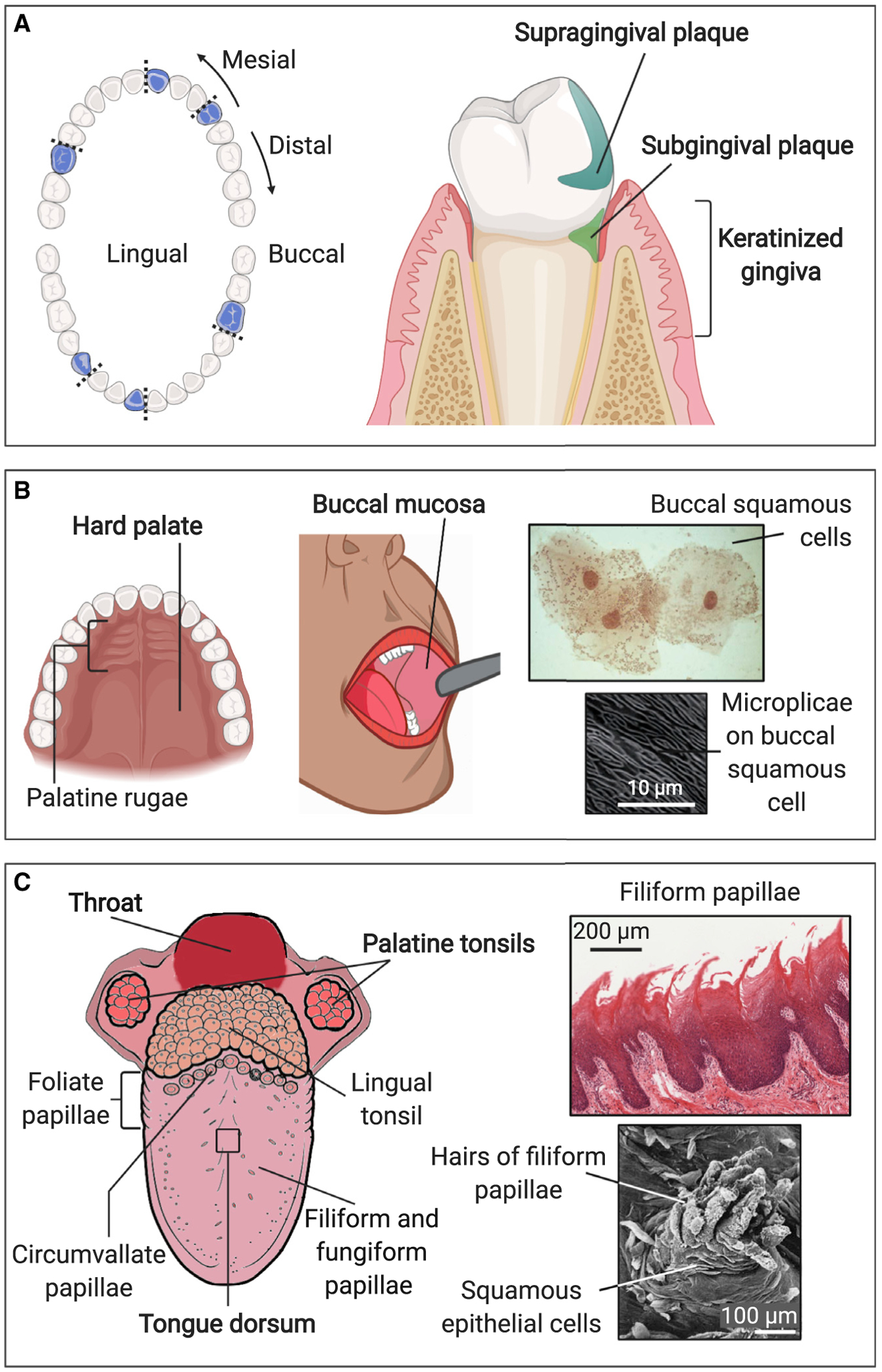
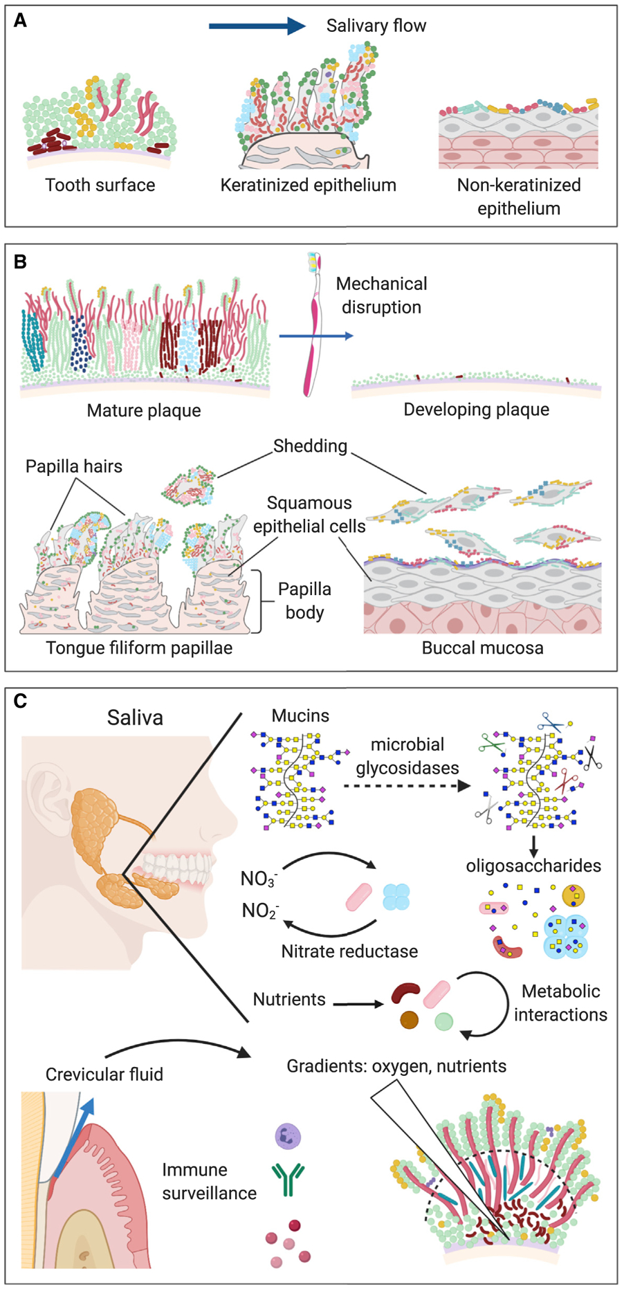
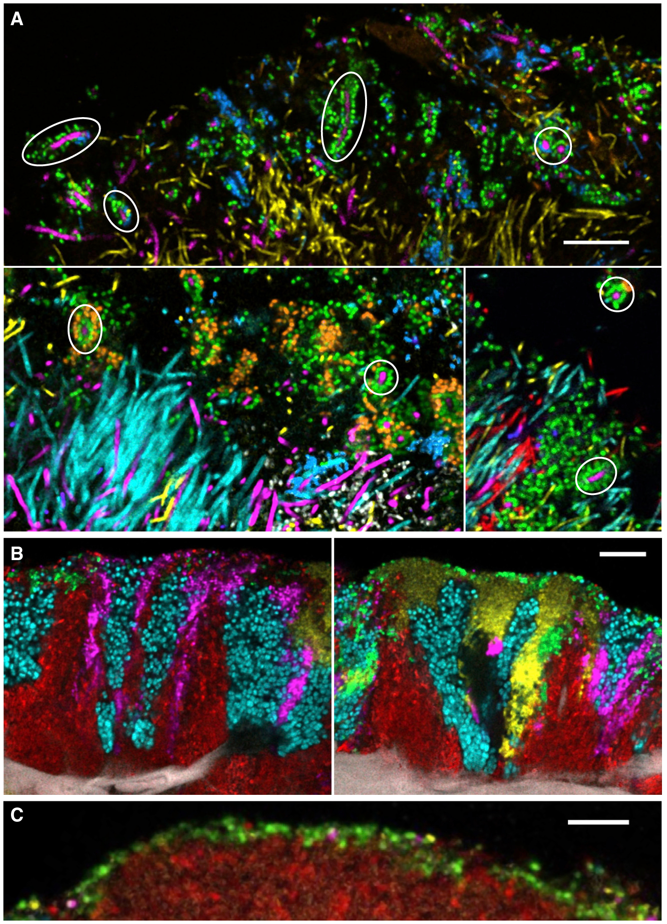
The mouth presents a multiplicity of local environments in communication with one another via saliva. The spatial organization of microbes within the mouth is shaped by opposing forces in dynamic equilibrium-salivary flow and adhesion, shedding and colonization-and by interactions among and between microbes and the host. Here we review recent evidence confirming that oral microbes are specialized for individual habitats within the mouth and that microbial habitats and niches are defined by micron-scale gradients in combination with short- and long-range interactions. Micron-scale structure illuminates the roles of individual taxa and provides insight into their community ecology and potential pathogenicity.


Figures

Figure 1.
Figure 1.. Major Habitats within the Mouth
The mouth contains a range of topographies and substrates that present varied habitats for microbes. Samples for DNA sequencing are usually collected by swabbing or scraping an area of 1 cm2 or more, but features at millimeter to micron scales could influence microbial colonization and create specialized micro-habitats supporting distinctive microbiotas. Sites sampled by the HMP are labeled in bold; some additional sites (not bold) are also shown. (A) Supragingival plaque, subgingival plaque, keratinized gingiva; blue indicates the position of teeth sampled in the HMP. (B) Hard palate, buccal mucosa; higher-magnification images show micro-habitats on buccal mucosa. Buccal mucosa smear modified from Datar et al. (2013), copyright 2013 Karger Publishers, Basel, Switzerland. Microplicae modified from Kullaa et al. (2014), reprinted by permission of the publisher, Taylor & Francis Ltd, http://www.tandfonline.com. (C) Palatine tonsils, throat, tongue dorsum; higher-magnification images show micro-habitats on tongue dorsum. Tongue diagram modified from Rice University’s OpenStax (https://openstax.org/books/anatomy-and-physiology/pages/23-3-the-mouth-pharynx-and-esophagus) under a Creative Commons International license (https://creativecommons.org/licenses/by/4.0); filiform papillae modified from https://histology.medicine.umich.edu/ - slide 116 under a Creative Commons Noncommercial-Share Alike license (https://creativecommons.org/licenses/by-nc-sa/3.0); hairs of filiform papillae modified from Kullaa-Mikkonen and Sorvari, 1985, copyright 1985 Karger Publishers, Basel, Switzerland. Illustrations created with BioRender.com.
Figure 2.
Figure 2.. Selective Forces within the Mouth
The oral microbiome is shaped by a dynamic equilibrium of opposing factors: flow and adhesion, shedding and colonization, and host and microbe. (A) Flow and adhesion. Salivary flow exerts a selective requirement for adhesion to oral substrates, both for retention in the mouth and for localization to a favorable metabolic environment. Distinctive oral substrates include the tooth surface, keratinized epithelia, and non-keratinized epithelia. (B) Shedding and colonization. Mechanical disruption of the biofilm or shedding of the underlying substrate is balanced by colonization of newly available substrate. The development of distinctive communities at the different oral sites occurs by differential binding of microbes to the different oral substrates, regrowth of the residual microbial biofilm, and colonization of the growing biofilm by additional taxa. Long-lived surfaces such as tooth surface and tongue dorsum develop thick biofilms, whereas the biofilm on rapidly shedding surfaces such as buccal mucosa is thin. (C) Host and microbe. The host and the microbial community exert mutual reciprocal influences on each other through binding interactions, immune surveillance, and gradients of nutrients and solutes. The host secretes salivary mucins, which are complex glycoproteins that support the growth of mixed syntrophic communities of microbes that possess glycosidases capable of releasing oligosaccharides from mucins. Secretion of nitrate and other nutrients into saliva by the host, and release of crevicular fluid from the gingival crevice into the mouth, could also serve to foster the growth of particular microbes, while immune surveillance limits the growth of others. Microbial metabolism, in turn, can generate strong localized gradients of oxygen and nutrients. The positioning of microbes at favorable locations within these gradients can lead to metabolic interaction and spatial structure within the microbial community. Illustrations created with BioRender.com.
Figure 3.
Figure 3.. Diversity of Micron-Scale Associations in the Oral Microbiome
(A) Some microbial taxa have highly specific structural associations. Corncobs in dental plaque are characterized by specific binding of Streptococcus sp. (green) or Porphyromonas sp. (blue) to Corynebacterium sp. (magenta), even when other filamentous taxa such as Fusobacterium (yellow), Leptotrichia (cyan), and Capnocytophaga (red) are present and apparently available for binding in the same area. Circles indicate examples of corncobs formed around Corynebacterium filaments. Scale bar, 10 μm. Modified from Mark Welch et al. (2016). (B) Some microbial taxa tend to form clonal clusters. The five taxa visualized here on the tongue dorsum occur in patches that have different characteristic shapes and relations to the underlying host epithelial cells but show clonal boundary relationships with each of the other 4 taxa. For example, Rothia (cyan) can be found next to Actinomyces (red), Neisseriaceae (yellow), Veillonella (magenta), and Streptococcus (green). Scale bar, 10 μm. Image credit: Steven Wilbert. (C) Streptococcus (green) often forms a thin (one-two cell thick) layer at the periphery of a tongue dorsum consortium—in this case, over a cluster of Actinomyces (red). Scale bar, 10 μm. Modified from Wilbert et al. (2020).

References

    1. Bowen WH, Burne RA, Wu H, and Koo H (2018). Oral Biofilms: Pathogens, Matrix, and Polymicrobial Interactions in Microenvironments. Trends Microbiol. 26, 229–242. - PMC - PubMed
    1. Carpenter GH (2020). Salivary Factors that Maintain the Normal Oral Commensal Microflora. J. Dent. Res 99, 644–649. - PMC - PubMed
    1. Datar U, Angadi PV, Hallikerimath S, and Kale AD (2013). Cytological assessment of Barr bodies using aceto-orcein and papanicolaou stains in buccal mucosal smears and their sex estimation efficacy in an Indian sample. Acta Cytol. 57, 516–521. - PubMed
    1. Dawes C (2003). Estimates, from salivary analyses, of the turnover time of the oral mucosal epithelium in humans and the number of bacteria in an edentulous mouth. Arch. Oral Biol 48, 329–336. - PubMed
    1. Dewhirst FE, Chen T, Izard J, Paster BJ, Tanner ACR, Yu W-H, Lakshmanan A, and Wade WG (2010). The human oral microbiome. J. Bacteriol 192, 5002–5017. - PMC - PubMed

Publication types

LinkOut - more resources