. 2020 Nov;98(5):1296-1307.
doi: 10.1016/j.kint.2020.07.019. Epub 2020 Aug 10.

SARS-CoV-2 causes a specific dysfunction of the kidney proximal tubule

Collaborators, Affiliations

SARS-CoV-2 causes a specific dysfunction of the kidney proximal tubule

Alexis Werion et al. Kidney Int. 2020 Nov.

Abstract

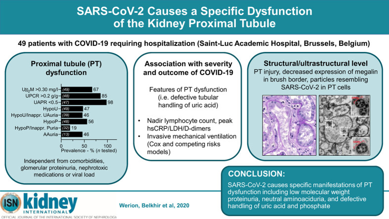
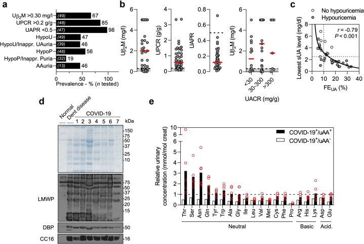
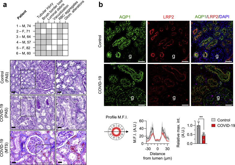
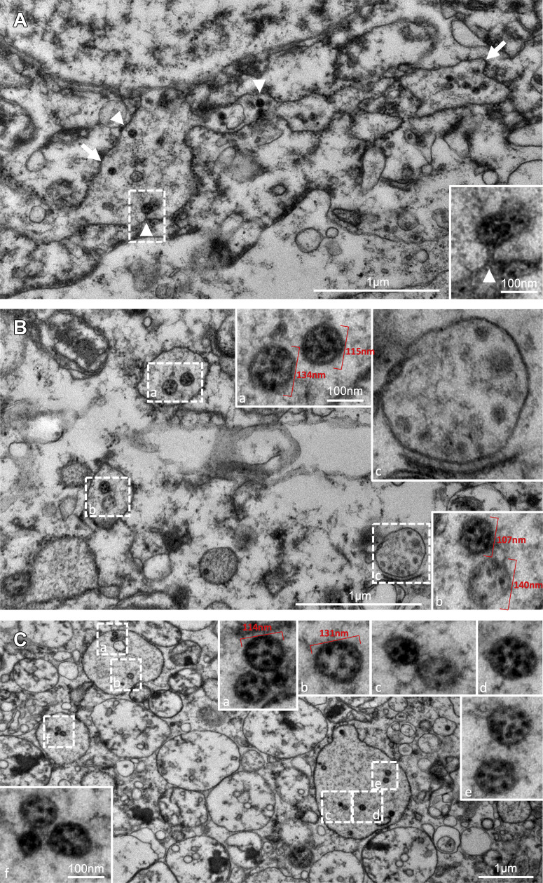
Coronavirus disease 2019 (COVID-19) is commonly associated with kidney damage, and the angiotensin converting enzyme 2 (ACE2) receptor for SARS-CoV-2 is highly expressed in the proximal tubule cells. Whether patients with COVID-19 present specific manifestations of proximal tubule dysfunction remains unknown. To test this, we examined a cohort of 49 patients requiring hospitalization in a large academic hospital in Brussels, Belgium. There was evidence of proximal tubule dysfunction in a subset of patients with COVID-19, as attested by low-molecular-weight proteinuria (70-80%), neutral aminoaciduria (46%), and defective handling of uric acid (46%) or phosphate (19%). None of the patients had normoglycemic glucosuria. Proximal tubule dysfunction was independent of pre-existing comorbidities, glomerular proteinuria, nephrotoxic medications or viral load. At the structural level, kidneys from patients with COVID-19 showed prominent tubular injury, including in the initial part of the proximal tubule, with brush border loss, acute tubular necrosis, intraluminal debris, and a marked decrease in the expression of megalin in the brush border. Transmission electron microscopy identified particles resembling coronaviruses in vacuoles or cisternae of the endoplasmic reticulum in proximal tubule cells. Among features of proximal tubule dysfunction, hypouricemia with inappropriate uricosuria was independently associated with disease severity and with a significant increase in the risk of respiratory failure requiring invasive mechanical ventilation using Cox (adjusted hazard ratio 6.2, 95% CI 1.9-20.1) or competing risks (adjusted sub-distribution hazard ratio 12.1, 95% CI 2.7-55.4) survival models. Thus, our data establish that SARS-CoV-2 causes specific manifestations of proximal tubule dysfunction and provide novel insights into COVID-19 severity and outcome.

Keywords: COVID-19; kidney; renal Fanconi syndrome; severe acute respiratory syndrome.


Figures

None
Graphical abstract
Figure 1
Figure 1
Proximal tubule dysfunction in patients with coronavirus disease 2019 (COVID-19). (a) Prevalence (%) of signs of proximal tubule (PT) dysfunction in patients with active COVID-19. Numbers (n) of patients tested are shown in brackets inside the bars. (b) Distribution of urinary β2-microglobulin (Uβ2M), urinary protein-to-creatinine ratio (UPCR), and urinary albumin-to-protein ratio (UAPR), and relationship between Uβ2M and urinary albumin-to-creatinine ratio in patients with COVID-19. Circles represent individual values, and red lines represent medians. (c) Inverse relationship between the lowest serum uric acid (sUA) level and fractional excretion of uric acid (FEUA) in patients with (gray circles) versus without (open circles) hypouricemia. Spearman’s coefficient r = –0.79 (95% confidence interval –0.89, –0.63), P < 0.001, n = 39. (d) Sodium dodecyl sulfate polyacrylamide gel electrophoresis followed by Coomassie blue staining and immunoblot shows the presence of low-molecular weight proteins (LMWP), vitamin D binding protein (DBP), and Clara cell secretory protein (CC16). Normal urine and urine from a patient with Dent disease (a rare inherited PT dysfunction caused by CLCN5 mutation) are shown as controls. Molecular weights (in kDa) of the urinary proteins are provided. (e) Relative urinary concentration of amino acids in patients with COVID-19 and aminoaciduria (COVID-19+/uAA+; black bars) or no aminoaciduria (COVID-19+/uAA-; open bars). The reference corresponds to the upper limit of normal. Individual values are represented by circles, and bars represent the mean values. AAuria; aminoaciduria; creat, creatinine; HypoP, hypophosphatemia; HypoU, hypouricemia; inappr. Puria, inappropriate phosphaturia; inappr. UAuria, inappropriate uricosuria. Ala, alanine; Arg, arginine; Asn, asparagine; Asp, aspartate; Cys, cysteine; Gln, glutamine; Glu, glutamate; Gly, glycine; His, histidine; Ile, isoleucine; Leu, leucine; Lys, lysine; Met, methionine; Phe, phenylalanine; Pro, proline; Ser, serine; Thr, threonine; Trp, tryptophan; Tyr, tyrosine; Val, valine. To optimize viewing of this image, please see the online version of this article at www.kidney-international.org.
Figure 2
Figure 2
Proximal tubule (PT) damage in kidneys of coronavirus disease 2019 (COVID-19) patients. (a) Pathologic scoring of kidney lesions in COVID-19. The grid summarizes pathologic findings on postmortem examination in 6 patients with COVID-19 and the presence (gray) or absence (white) of tubular injury, brush border loss, debris in the lumen, red blood cell (RBC) aggregates, vacuolization of tubular cells, and glomerular (glom.) alterations. Representative pictures of sections from control and COVID-19 kidneys, stained with periodic acid–Schiff (PAS) or Masson’s trichrome staining (MTS), show prominent tubular lesions with dilation, mitoses in epithelial cells, major alterations of the brush border (i.e., in the S1 segment of the PT), shedding of cellular debris into the lumen, as well as erythrocytes aggregates in peritubular capillaries. Original magnification ×20 and ×40; bars = 50 μm and 25 μm, on left and right pictures, respectively. (b) Representative pictures of double immunostaining with anti-aquaporin (AQP) 1 (green channel) and anti-megalin (low-density lipoprotein receptor–related protein [LRP] 2, red channel) antibodies viewed under confocal fluorescence microscopy in kidney sections from a control and a patient with COVID-19. Nuclei are stained with 4′,6-diamidino-2-phenylindole (DAPI; blue channel). Original magnification ×20. Bars = 50 μm. Mean fluorescence intensity (M.F.I.) profiles and relative maximal intensity (max. int.) for LRP2 were quantified on cross sectional sections of PT from 3 controls (gray) and 5 patients who died of COVID-19 (red). Data are mean values and SEM. P = 0.002, unpaired t test. M, male; F, female. To optimize viewing of this image, please see the online version of this article at www.kidney-international.org.
Figure 3
Figure 3
Transmission electron microscopy of particles in proximal tubule cells in a kidney sample of an autopsied patient deceased from coronavirus disease 2019 (COVID-19). (A) Low-magnification electron microscopy image showing numerous particles of approximately 90–140 nm in diameter within the lumen of rough endoplasmic reticulum (RER) (arrows), some of which have budded into the lumen from the cytoplasm (arrowheads). Inset (a) shows the budding and the stalk of a particle. Size is indicated by the magnification bars. (B) Particles observed inside vacuoles formed by RER and lighter densities inside smooth endoplasmic reticulum. Areas seen in the lower magnification marked by dashed-line boxes are enlarged in solid-line insets and are labeled correspondingly. Insets (a) and (b) are representative of numerous particles containing dense smudgy dots in similarly sized particles observed within RER elsewhere. In some particles (inset a), the trilaminar membrane envelope is evident. A larger vacuole (inset c) without ribosomes studding the outside and located near the Golgi apparatus contains irregularly sized densities and does not contain viruses. Size of the low magnification is indicated on the print; size in the high magnifications is the same in all insets and is indicated by the bar in inset (a). (C) Particles shown (similarly to those in B above) at low magnification in the large image in dashed-line boxes and at high magnification in solid-line insets corresponding to the dashed-line boxes (a–f). All particles have irregular dense black dots inside, and a few (e.g., see insets b and e) have crisp small rings, possibly exact cross sections of tubular or helical structures. Size of the low magnification is indicated on the print; size in the high magnifications is the same in all insets and is indicated by the bar in inset (f). To optimize viewing of this image, please see the online version of this article at www.kidney-international.org.

Comment in

References

    1. Acter T., Uddin N., Das J. Evolution of severe acute respiratory syndrome coronavirus 2 (SARS-CoV-2) as coronavirus disease 2019 (COVID-19) pandemic: a global health emergency. Sci Total Environ. 2020;730:138996. - PMC - PubMed
    1. Wu Z., McGoogan J.M. Characteristics of and important lessons from the Coronavirus Disease 2019 (COVID-19) outbreak in China: summary of a report of 72 314 cases from the Chinese Center for Disease Control and Prevention. JAMA. 2020;323:1239–1242. - PubMed
    1. Guan W.J., Ni Z.Y., Hu Y. Clinical characteristics of Coronavirus Disease 2019 in China. N Engl J Med. 2020;382:1708–1720. - PMC - PubMed
    1. Li W., Moore M.J., Vasilieva N. Angiotensin-converting enzyme 2 is a functional receptor for the SARS coronavirus. Nature. 2003;426:450–454. - PMC - PubMed
    1. Hamming I., Timens W., Bulthuis M.L. Tissue distribution of ACE2 protein, the functional receptor for SARS coronavirus. A first step in understanding SARS pathogenesis. J Pathol. 2004;203:631–637. - PMC - PubMed

Publication types

MeSH terms

LinkOut - more resources