. 2020 Jul 24:11:652.
doi: 10.3389/fneur.2020.00652. eCollection 2020.

Improving Function in Cavernous Sinus Meningiomas: A Modern Treatment Algorithm

Affiliations

Improving Function in Cavernous Sinus Meningiomas: A Modern Treatment Algorithm

André Beer-Furlan et al. Front Neurol. .

Abstract

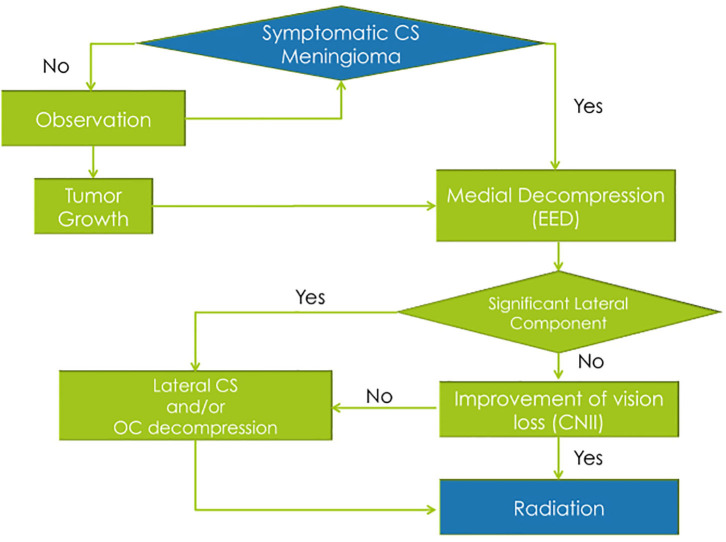
Background: The efficacy and safety of radiosurgery led to paradigm shift in the management of cavernous sinus meningiomas. Nevertheless, patients are still significantly affected by cranial nerve deficits related to the mass effect of these tumors. Our management strategy involves the combination of a functional surgical decompression followed by radiation therapy. Methods: We reviewed a single institution's cohort of patients who underwent endoscopic endonasal decompression (EED) for symptomatic meningiomas primarily involving the cavernous sinus (CS) from 2010 to 2016. The preoperative neuro-ophthalmological exam was compared to the 1- and 6-month postoperative exams. The patient's length of hospital stay, complications, and radiological and clinical follow-up were noted. Results: A total of 17 patients underwent EED for CS meningiomas that fit our radiological criteria. The final outcome at the 6-month visit showed five patients (62.5%) with normalization of deficit and three patients (37.5%) with partial improvement of the CNII deficit. Out of the 12 patients who had cavernous sinus cranial nerves (CSCN) deficits, the final outcome at the 6-month visit showed four patients (33.33%) with normalization of deficit, seven patients (58.3%) with partial improvement, and one patient (8.33%) with no improvement. There were no intraoperative complications. Conclusion: The EED for CS meningiomas is a valuable technique when addressing acute/subacute CNII and CSCN deficits. This conservative surgical approach showed good functional outcomes, low morbidity, and low complication rates. However, it does not exempt the need for radiosurgery/radiation therapy for control of tumor growth.

Keywords: cavernous sinus; endoscopic endonasal; meningioma; parasellar; skull base.


Figures

Figure 1
Figure 1
Calculation of the total tumor volume using computer software. (A,B) Axial T1 with gadolinium images; (C) coronal T1 with gadolinium image; (D) sagittal T1 with gadolinium image.
Figure 2
Figure 2
Calculation of the intracavernous volume of tumor using computer software. (A,B) Axial T1 with gadolinium images; (C) coronal T1 with gadolinium image; (D) sagittal T1 with gadolinium image.
Figure 3
Figure 3
Summary of the “functional skull base surgery” strategy used in cavernous sinus meningiomas.
Figure 4
Figure 4
Illustration of the surgical anatomy. (A) Endoscopic view showing the cavernous sinus anatomy after removal of the bone overlying the sella and cavernous sinus; (B) coronal cross-section illustration demonstrating the area of bone decompression.
Figure 5
Figure 5
Intraoperative images of endoscopic endonasal decompression for a right side CS meningioma. (A) Drilling down the intersphenoid septum and accessory septa flush with the ventral skull base; (B) exposure of the sellar dura and CS dura with a bridge of bone overlying the cavernous internal carotid artery; (C) complete decompression of the bone overlying the sella, medial SOF, and CS; (D) opening the sellar interdural space (between the periosteal and meningeal layers of dura) that is occupied by the tumor and it is considered one of the safe zones for biopsy. CS, cavernous sinus; SOF, superior orbital fissure; V2, maxillary nerve.
Figure 6
Figure 6
Intraoperative images of endoscopic endonasal decompression for a left side CS meningioma. (A) Drilling down the hyperostotic bone overlying the CS; (B) exposure of the sellar dura; (C) opening the sellar interdural space after complete decompression; (D) decompression of bone overlying the sella, medial SOF, and CS. CS, cavernous sinus; SOF, superior orbital fissure; V2, maxillary nerve.
Figure 7
Figure 7
(A) Preoperative coronal CTA images (bone window) of a patient with a right side cavernous sinus meningioma; (B) postoperative coronal CT images (bone window) showing the area of bone decompression on the sella, medial aspect of the superior orbital fissure and cavernous sinus.

References

    1. DeMonte F, Smith HK, Al-Mefty O. Outcome of aggressive removal of cavernous sinus meningiomas. J Neurosurg. (1994) 81:245–51. 10.3171/jns.1994.81.2.0245 - DOI - PubMed
    1. O'Sullivan MG, van Loveren HR, Tew JM. The surgical resectability of meningiomas of the cavernous sinus. Neurosurgery. (1997) 40:238–44. 10.1097/00006123-199702000-00003 - DOI - PubMed
    1. Correa SFM, Marta GN, Teixeira MJ. Neurosymptomatic carvenous sinus meningioma: a 15-years experience with fractionated stereotactic radiotherapy and radiosurgery. Radiat Oncol. (2014) 9:27. 10.1186/1748-717X-9-27 - DOI - PMC - PubMed
    1. Park K-J, Kano H, Iyer A, Liu X, Tonetti DA, Lehocky C, et al. . Gamma knife stereotactic radiosurgery for cavernous sinus meningioma: long-term follow-up in 200 patients. J Neurosurg. (2018) 1:1–10. 10.3171/2018.2.JNS172361 - DOI - PubMed
    1. Klinger DR, Flores BC, Lewis JJ, Barnett SL. The treatment of cavernous sinus meningiomas: evolution of a modern approach. Neurosurg Focus. (2013) 35:E8. 10.3171/2013.9.FOCUS13345 - DOI - PubMed

LinkOut - more resources