. 2020 Jul 10:11:443.
doi: 10.3389/fendo.2020.00443. eCollection 2020.

An ex vivo Approach to Study Hormonal Control of Spermatogenesis in the Teleost Oreochromis niloticus

Affiliations

An ex vivo Approach to Study Hormonal Control of Spermatogenesis in the Teleost Oreochromis niloticus

Michelle Thönnes et al. Front Endocrinol (Lausanne). .

Abstract

As the male reproductive organ, the main task of the testis is the production of fertile, haploid spermatozoa. This process, named spermatogenesis, starts with spermatogonial stem cells, which undergo a species-specific number of mitotic divisions until starting meiosis and further morphological maturation. The pituitary gonadotropins, luteinizing hormone, and follicle stimulating hormone, are indispensable for vertebrate spermatogenesis, but we are still far from fully understanding the complex regulatory networks involved in this process. Therefore, we developed an ex vivo testis cultivation system which allows evaluating the occurring changes in histology and gene expression. The experimental circulatory flow-through setup described in this work provides the possibility to study the function of the male tilapia gonads on a cellular and transcriptional level for at least 7 days. After 1 week of culture, tilapia testis slices kept their structure and all stages of spermatogenesis could be detected histologically. Without pituitary extract (tilPE) however, fibrotic structures appeared, whereas addition of tilPE preserved spermatogenic cysts and somatic interstitium completely. We could show that tilPE has a stimulatory effect on spermatogonia proliferation in our culture system. In the presence of tilPE or hCG, the gene expression of steroidogenesis related genes (cyp11b2 and stAR2) were notably increased. Other testicular genes like piwil1, amh, or dmrt1 were not expressed differentially in the presence or absence of gonadotropins or gonadotropin containing tilPE. We established a suitable system for studying tilapia spermatogenesis ex vivo with promise for future applications.

Keywords: Amh (anti-Müllerian hormone); FSH; LH; Nile tilapia (Oreochromis niloticus); hCG; pituitary extract; spermatogenesis; testis culture.


Figures

Figure 1
Figure 1
Confocal images of vibratome sections of testis explants cultured for 7 days in a flow-through chamber and activated by pituitary extracts. (A) Picture of complete flow-through culture system; 1—temperature unit, 2—chambers, 3—peristaltic pump, 4—media bottles, 5—connecting tubes with sterile filter in the backflow. (B) Chamber top view, 1—inflow, 2—vent, 3—medium filled part of the chamber, 4—outflow. (C) Chamber bottom view, white arrows mark the access channels to the in- and outflow exemplarily (3 out of 10). (D) Intact testis morphology with spermatogenic tubules (dashed lines), proliferating germ line cells in cysts (red label by Pcna antibody), single Pcna positive type A spermatogonia (arrows), well developed interstitium with Leydig cell groups (*), tunica albuginea (TA); DNA (Draq5 in false color)—blue, for visualization of cellular size and shape β-catenin antibody (green) was used—preferably type A spermatogonia and Leydig cells were stained. (E) Many Pcna positive germ line cells in cysts (exemplary labeled by a dotted line) and single type A spermatogonia (arrows). (F) Pcna-positive, undifferentiated type A spermatogonia (identification by nuclear morphology, arrow) adjacent to a Pcna positive Sertoli cell (SE). (G) Cyst with Pcna expressing type B spermatogonia (SgB). Scale bars: 20 μm in (D,E), 10 μm in (F,G).
Figure 2
Figure 2
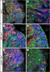
Confocal images of Pcna (red) and β-catenin (green) immunolabeled vibratome sections of 7 days cultivated testis explants. (A,B) Testis sections from two different explants from the beginning of the flow-through culture (day 0). Large groups of Leydig cells are indicated by an asterisk (*). Nucleated erythrocytes (exemplary labeled with “ery”) inside of these groups appear in a light gray-blue coloration (shown in higher magnification in Figure S3). Spermatogenic tubules contain cysts with cells of all stages of spermatogenesis. Abundant Pcna labeling of “single” SgA (exemplarily shown by a white arrow) and cyst consisting of SgB and spermatocytes occurred. Postmeiotic germ line cells show no Pcna and appear in blue by Draq5 stained nuclei inside of the tubules. Some spermatogenic tubules are labeled by a white dashed line exemplarily. (C,D) Testis sections from two different explants show completely preserved testis tissue after 7 days of flow-through culture with tilPE supplementation. Many Pcna labeled cyst cells, Pcna labeled type A spermatogonia and large groups of Leydig cells (*) are present. The interstitium appears unchanged in comparison to the start of the culture at day 0. (E,F) Testis sections from two different explants after 7 days of flow-through culture without addition of tilPE (control). Normally developed spermatogenic tubules are present. Cysts with all stages of spermatogenesis and Pcna labeling occur (E). In a lot of germ cell cysts an unusual cytoplasmic Pcna localization can be seen (exemplarily labeled by flat triangles). The structure of the interstitium has changed, large Leydig cells groups are not obvious and fibrotic structures appear (exemplarily labeled by a diamond #). An example of severe fibrosis is shown in (F). Here, a lot of interstitial cells are Pcna labeled and β-catenin staining differs from the mesh-like pattern of caused by the presence of Leydig cells. Remaining germ line cysts of the tubules display spermatids (St) or very early germ line cells (SgA—white arrow) only. The proportion of type B spermatogonia and spermatocytes seems to be reduced in (E) and these cell types have disappeared in (F). A morphological comparison in higher magnification is shown in Figure S2 and further details in Figures S4, S5. In all pictures DNA is stained by Draq5 (in false color as blue). Scale bars: 50 μm.
Figure 3
Figure 3
Evidence for spermatogonia proliferation. (A) The relative number of SgA and SgB after 7 days of organ culture. The ratio between SgA and SgB does not significantly change between the treatments. (B) Ratio of EdU positive spermatogonia. Compared to the control, more SgB had incorporated EdU after 7 days. EdU was added for the last 24 h of the culture. X2 test was applied: ***p < 0.001. The data displayed in (A,B) came from two independent experiments. Testis sections from 3 male fish were analyzed in total. (C) Representative testis section for spermatogonia counting (example from a 7-day culture without tilPE supplementation). Nuclei (gray) are stained with DAPI. Spermatogonia type A (white arrows) and B (SgB) were classified after nuclear diameter and Vasa expression (green). EdU incorporation (red) was used to detect proliferating cells. Sertoli cells were exemplary labeled with SE. (D) Green channel only of picture (C) to illustrate the Vasa positive cells and different intensities of the Vasa signals. Scale bar: 10 μm.
Figure 4
Figure 4
Gene expression after 7 days of organ culture. The gene expression is given as fold change compared to the untreated control. Data are represented as average fold change and SEM. tilPE treatment data includes 4 independent experiments, hCG and tilPE + hCG data include 3 independent experiments. Gene expression was measured with qPCR in three technical replicates per pooled sample. A two tailed Wilcoxon rank-sum test was used to test for significant differences between treatment and control. Significance levels were interpreted as follows: *p < 0.05, and ***p < 0.001.

Similar articles

Cited by

References

    1. Devlin RH, Nagahama Y. Sex determination and sex differentiation in fish: an overview of genetic, physiological, and environmental influences. Aquaculture. (2002) 208:191–364. 10.1016/S0044-8486(02)00057-1 - DOI
    1. Schulz RW, de França LR, Lareyre JJ, LeGac F, Chiarini-Garcia H, Nobrega RH, et al. . Spermatogenesis in fish. Gen Comp Endocrinol. (2010) 165:390–411. 10.1016/j.ygcen.2009.02.013 - DOI - PubMed
    1. Chauvigné F, Zapater C, Gasol JM, Cerdà J. Germ-line activation of the luteinizing hormone receptor directly drives spermiogenesis in a nonmammalian vertebrate. Proc Natl Acad Sci USA. (2014) 111:1427–32. 10.1073/pnas.1317838111 - DOI - PMC - PubMed
    1. Schulz RW, Menting S, Bogerd J, França LR, Vilela DAR, Godinho HP. Sertoli cell proliferation in the adult testis—evidence from two fish species belonging to different orders. Biol Reprod. (2005) 73:891–8. 10.1095/biolreprod.105.039891 - DOI - PubMed
    1. França LR, Nóbrega RH, Morais RDVS, De Castro Assis LH, Schulz RW. Sertoli cell structure and function in anamniote vertebrates. Sertoli Cell Biol. (2015) 385–407. 10.1016/b978-0-12-417047-6.00013-2 - DOI

Publication types