

Delayed cardiac tamponade following blunt chest trauma due to disruption of fourth costal cartilage with posterior dislocation
- PMID: 32793793
- PMCID: PMC7415922
- DOI: 10.1016/j.tcr.2020.100340
Delayed cardiac tamponade following blunt chest trauma due to disruption of fourth costal cartilage with posterior dislocation
Erratum in
-
Erratum regarding missing patient consent statement in previously published articles.Trauma Case Rep. 2023 Mar 1;45:100808. doi: 10.1016/j.tcr.2023.100808. eCollection 2023 Jun. Trauma Case Rep. 2023. PMID: 37197575 Free PMC article.
Abstract
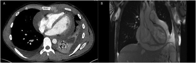
Cardiac tamponade is a recognised sequelae of non-penetrating and penetrating chest trauma. Delayed cardiac tamponade has been described following blunt chest trauma. We present a 29 year-old gentleman who had initially presented to peripheral district general hospital following direct blunt chest wall trauma. His initial trauma CT demonstrated a small mediastinal haematoma and large left haemopneumothorax and disruption/dislocation of the costal cartilage. He initially underwent a thoracoscopic procedure uneventfully. He then had worsening chest radiograph appearances with enlarging cardiac contours. Transthoracic echocardiography confirmed cardiac tamponade. He underwent creation of a pericardial window and excision of the protruding fourth costal cartilage.
Keywords: Cardiac tamponade; Costal cartilage; Haemothorax; Pneumothorax; Thoracoscopy.
Crown Copyright © 2020 Published by Elsevier Ltd.
Conflict of interest statement
None to declare.
Figures

References
-
- Hermens J.A., Wajon E.M., Grandjean J.G., Haalebos M.M., von Birgelen C. Delayed cardiac tamponade in a patient with previous minor blunt chest trauma. Int. J. Cardiol. 2009;131(3):124–126. 24. - PubMed
-
- El-Menyar A., Al Thani H., Zarour A., Latifi R. Understanding traumatic blunt cardiac injury. Ann. Card. Anaesth. 2012;15:287–295. - PubMed
Publication types
LinkOut - more resources
Full Text Sources
