Review
. 2020 Aug 20;27(8):904-920.
doi: 10.1016/j.chembiol.2020.07.022. Epub 2020 Aug 13.

Seeing (and Using) the Light: Recent Developments in Bioluminescence Technology

Affiliations
Review

Seeing (and Using) the Light: Recent Developments in Bioluminescence Technology

Anna C Love et al. Cell Chem Biol. .

Abstract

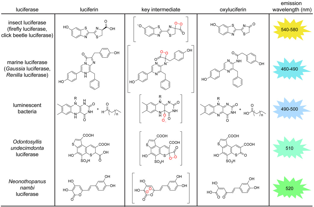
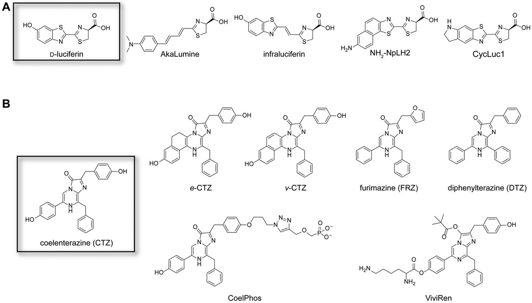
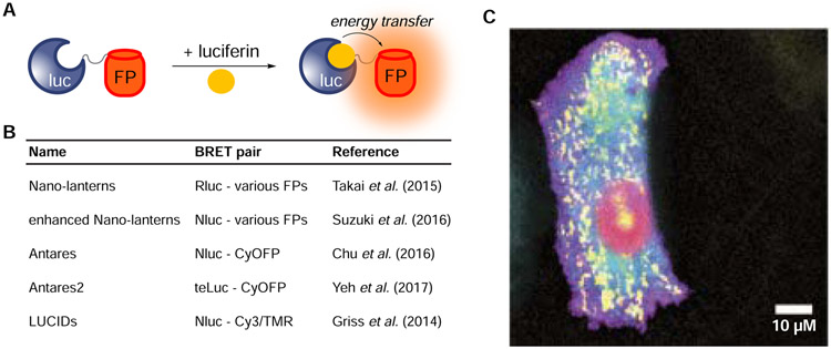
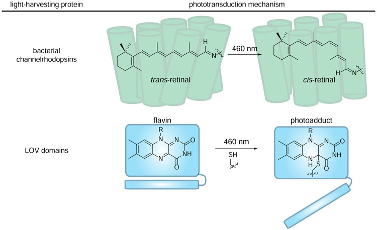
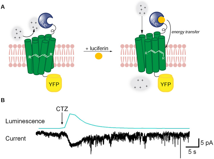
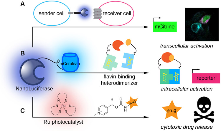
Bioluminescence has long been used to image biological processes in vivo. This technology features luciferase enzymes and luciferin small molecules that produce visible light. Bioluminescent photons can be detected in tissues and live organisms, enabling sensitive and noninvasive readouts on physiological function. Traditional applications have focused on tracking cells and gene expression patterns, but new probes are pushing the frontiers of what can be visualized. The past few years have also seen the merger of bioluminescence with optogenetic platforms. Luciferase-luciferin reactions can drive light-activatable proteins, ultimately triggering signal transduction and other downstream events. This review highlights these and other recent advances in bioluminescence technology, with an emphasis on tool development. We showcase how new luciferins and engineered luciferases are expanding the scope of optical imaging. We also highlight how bioluminescent systems are being leveraged not just for sensing-but also controlling-biological processes.

Keywords: bioluminescence; imaging; luciferase; luciferin; optogenetics.


Figures

Figure 1.
Figure 1.. Bioluminescence in the wild.
Examples of naturally occurring luciferase enzymes and luciferin small molecules. All luciferases oxidize their complementary luciferins to yield photons of light, although unique chemistries are employed. Most bioluminescent probes emit light in the blue-green region. (*Emission wavelengths reported at room temperature.)
Figure 2.
Figure 2.. Noninvasive imaging in vivo with bioluminescent probes.
Luciferase-luciferin pairs have been routinely used to track cell migration and proliferation in vivo. (A) Imaging cancer cell proliferation and metastases. Mice were injected with Fluc-expressing MDA-MB-231 cells. Metastatic cells were harvested from lungs and re-injected into recipient mice. These cells exhibited a higher degree of metastatic outgrowth than the parental tumor. Figure reprinted with permission from Minn, et al., 2005 (B) Imaging T cell trafficking. Gluc-expressing T cells were injected into immunodeficient mice. T cell proliferation and homing were visualized over time via bioluminescence. Figure reprinted with permission from Santos et al., 2009.
Figure 3.
Figure 3.. Synthetic luciferins.
(A) Representative analogs of D-luciferin. Some of these molecules exhibit preferential reactivity with mutant luciferases, red-shifted emission, and/or improved bioavailability in organisms. (B) Representative analogs of coelenterazine. Some of these molecules exhibit unique reactivities with mutant luciferases, improved stability and solubility, and/or altered wavelengths of emission.
Figure 4.
Figure 4.. Multicolor imaging with engineered BRET reporters.
(A) A variety of luciferase-fluorescent protein (FP) fusions have been generated. Resonance energy transfer can provide a range of bioluminescent colors. (B) Example BRET reporters. These constructs comprise luciferase donors with either fluorescent protein or small molecule acceptors. (C) A panel of enhanced Nano- lanterns was expressed in HeLa cells, enabling real-time BRET imaging of cellular organelles. Reprinted with permission from Suzuki et al., 2016.
Figure 5.
Figure 5.. Light-harvesting proteins use unique chromophores to convert light energy to chemical energy.
(top) Channelrhodopsins comprise covalently bound retinal chromophores. Retinal isomerizes when illuminated with blue light, driving the channel pore to open. (bottom) LOV domains comprise noncovalently bound flavins chromophores. When excited with 460 nm light, flavin can be trapped by a nearby cysteine. The resulting conformational change initiates signal transduction.
Figure 6.
Figure 6.. Luciferase-rhodopsin fusions enable spatiotemporal control over ion channel function.
(A) The first-generation luminopsin (LMO1) featured Gluc (blue) tethered to channelrhodopsin2 (ChR2, green). CTZ application potentiates energy transfer, driving retinal isomerization and channel opening. (B) LMO1 was expressed in HEK cells. CTZ application generated photocurrents. Reprinted from Berglund, et al. 2013.
Figure 7.
Figure 7.. Recent examples of harnessing bioluminescent photons to drive biological processes.
(A) Transcellular activation of gene expression. Nluc expressed in “sender” cells (blue) activated light-sensitive proteins in “receiver” cells (pink), resulting in mCitrine expression (green). (B) Intracellular activation of gene expression. Energy transfer from a Nluc-mCerulean conjugate to a flavin-binding protein facilitated heterodimerization and downstream transcription of reporter genes. (C) Cytotoxic drug release. Energy transfer from Nluc to a ruthenium photocatalyst facilitated bond cleavage reactions and cargo release.

References

    1. Adams ST Jr, and Miller SC (2020). Enzymatic promiscuity and the evolution of bioluminescence. FEBS J. 287, 1369–1380. - PMC - PubMed
    1. Aird EJ, Tompkins KJ, Ramirez MP, and Gordon WR (2020). Enhanced molecular tension sensor based on bioluminescence resonance energy transfer (BRET). ACS Sens. 5, 34–39. - PMC - PubMed
    1. Andreu N, Zelmer A, Fletcher T, Elkington PT, Ward TH, Ripoll J, Parish T, Bancroft GJ, Schaible U, Robertson BD, Wiles S (2010). Optimisation of bioluminescent reporters for use with mycobacteria. PLoS one 5, e10777. - PMC - PubMed
    1. Atasoy D, and Sternson SM (2017). Chemogenetic tools for causal cellular and neuronal biology. Physiol. Rev 98, 391–418. - PMC - PubMed
    1. Azevedo MF, Nie CQ, Elsworth B, Charnaud SC, Sanders PR, Crabb BS, and Gilson PR (2014). Plasmodium falciparum transfected with ultra bright NanoLuc luciferase offers high sensitivity detection for the screening of growth and cellular trafficking inhibitors. PLoS One 9, e112571. - PMC - PubMed

Publication types