. 2021 Jan:221:299-310.
doi: 10.1016/j.ajo.2020.08.004. Epub 2020 Aug 12.

Sector Retinitis Pigmentosa: Extending the Molecular Genetics Basis and Elucidating the Natural History

Affiliations

Sector Retinitis Pigmentosa: Extending the Molecular Genetics Basis and Elucidating the Natural History

Michalis Georgiou et al. Am J Ophthalmol. 2021 Jan.

Abstract

Purpose: To determine the genetic background of sector retinitis pigmentosa (RP) natural history to better inform patient counseling.

Design: Retrospective case series.

Methods: Review of clinical notes, retinal imaging including color fundus photography (CFP), fundus autofluorescence (FAF), optical coherence tomography (OCT), electrophysiological assessment (ERG), and molecular genetic testing were performed in patients with sector RP from a single tertiary referral center. Main outcomes measured were demographic data, signs and symptoms, visual acuity, molecular genetics; and ERG, FAF, and OCT findings.

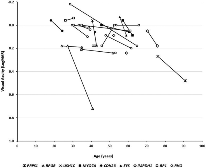
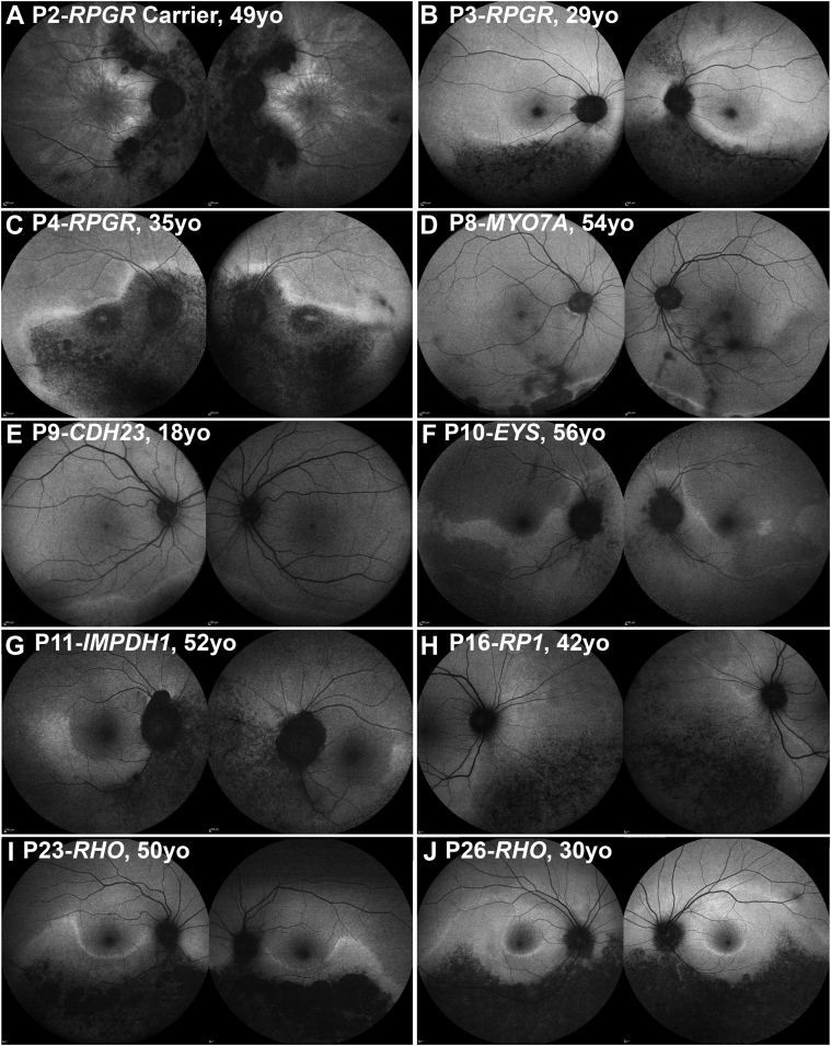
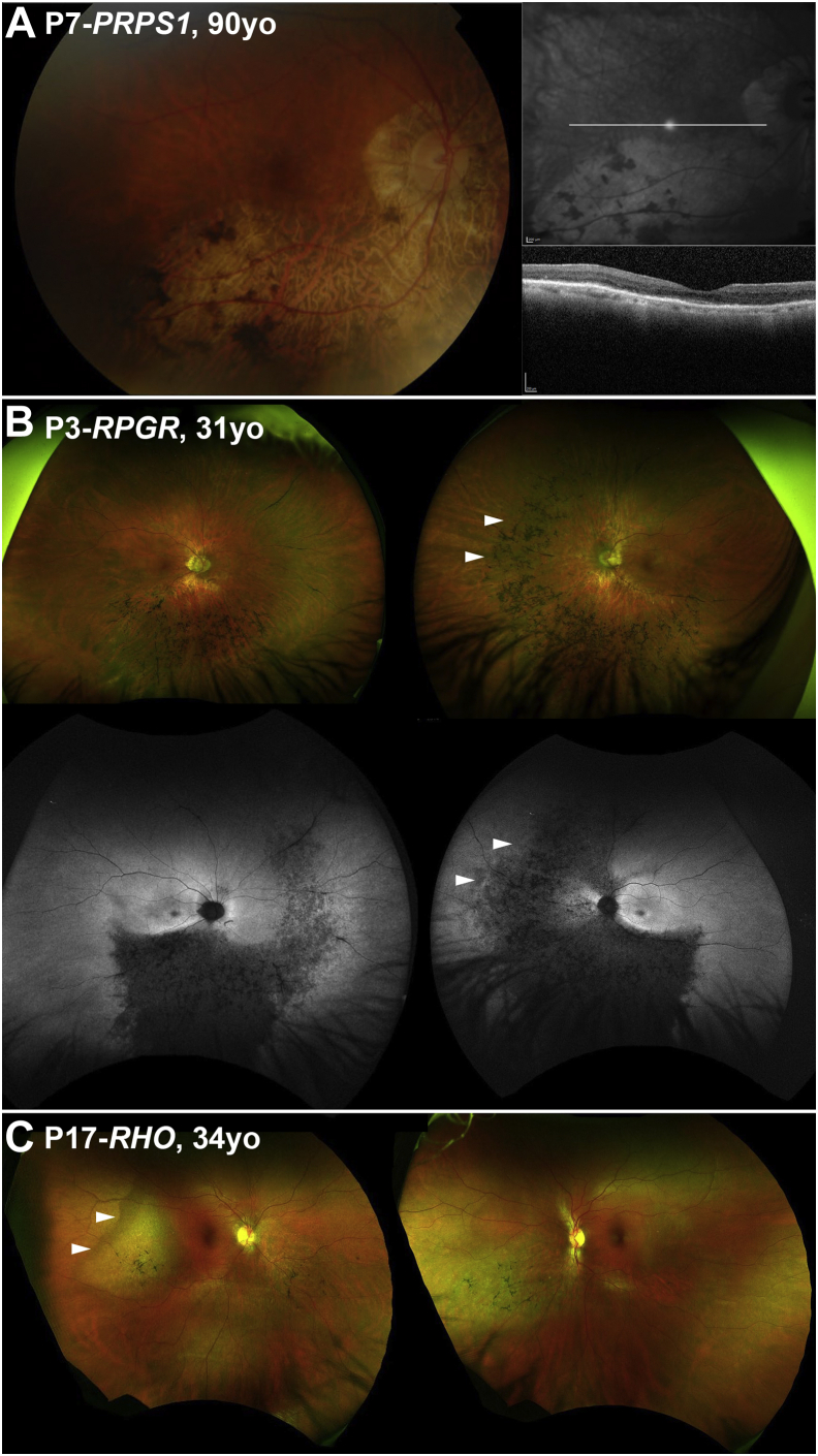
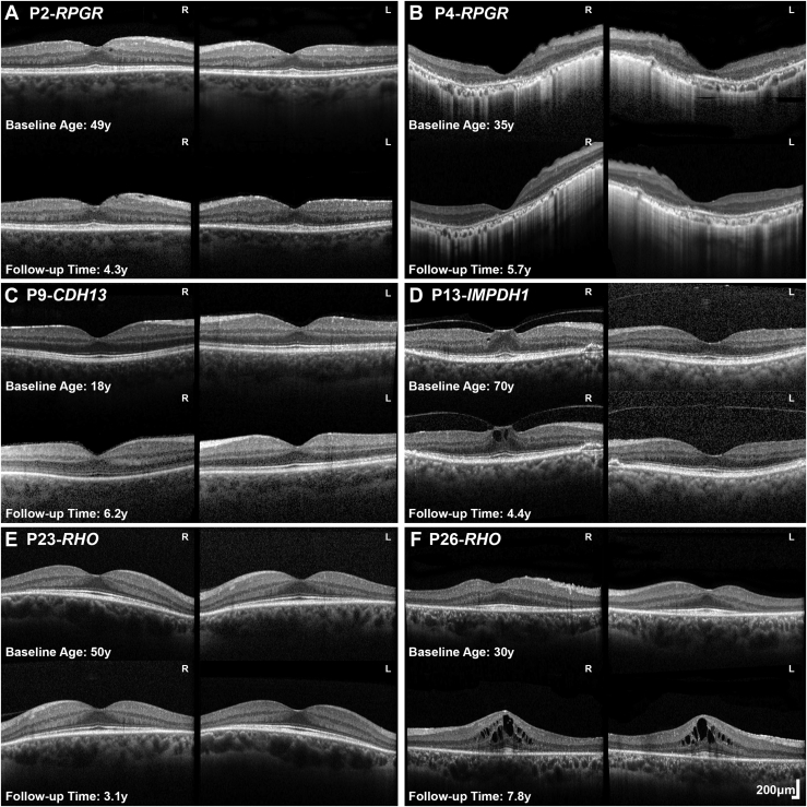
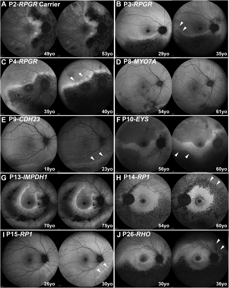
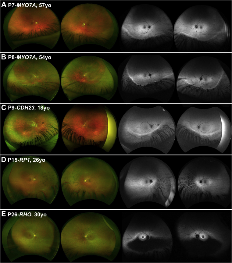
Results: Twenty-six molecularly confirmed patients from 23 different families were identified harboring likely disease-causing variants in 9 genes. The modes of inheritance were autosomal recessive (AR, n=6: USH1C, n=2; MYO7A, n=2; CDH3, n=1; EYS, n=1), X-linked (XL, n=4: PRPS1, n=1; RPGR, n=3), and autosomal dominant (AD, n=16: IMPDH1, n=3; RP1, n=3; RHO, n=10), with a mean age of disease onset of 38.5, 30.5, and 39.0 years old, respectively. Five of these genes have not previously been reported to cause sector RP (PRPS1, MYO7A, EYS, IMPDH1, and RP1). Inferior and nasal predilection was common across the different genotypes, and patients tended to maintain good central vision. Progression on serial FAF was observed in RPGR, MYO7A, CDH23, EYS, IMPDH1, RP1, and RHO-associated sector RP.

Conclusions: The genotypic spectrum of the disease is broader than previously reported. The longitudinal data provided will help to make accurate patient prognoses and counseling as well as inform patients' potential participation in the increasing numbers of trials of novel therapeutics and access to future treatments.


Figures

Figure 1
Figure 1
Visual acuity graphical representation. Twenty-four patients had available visual acuity data; 20 patients were longitudinally assessed and were followed for a mean of 10.6 years (range: 2.3-36.1 years). The graph represents visual acuity at baseline and follow-up (where available), over age. Each genotype is presented with a different marker.
Figure 2
Figure 2
Disease symmetry. Fundus autofluorescence imaging of the right and left eyes of 10 patients (A-J), with sector retinitis pigmentosa. The disease was symmetrical in all cases, except for P3 (B). The age and the genotype of everyone is noted in the figure.
Figure 3
Figure 3
Examples of disease asymmetry. (A) PRPS1 is known to cause asymmetrical disease in women. Multimodal imaging data were available only from the right eye precluding interocular comparison. (Left) Color fundus photograph. (Right) Near-infrared imaging of the right eye with the white line marking the location of the transfoveal optical coherence tomography scan presented below. (B and C) Examples of interocular asymmetry. (B) P3 has more advanced disease nasally in the left eye (white arrow heads), which are better visualized with fundus autofluorescence imaging (second line). (C) Color fundus photograph of P17, with more advanced disease temporal to the fovea in the right eye (white arrow heads).
Figure 4
Figure 4
Optical coherence tomography (OCT) in sector RP. (A-F) Transfoveal horizontal OCT scans of 6 patients with sector RP of both eyes at baseline and follow-up. Genotype, age at baseline and follow-up time are noted in the figure. (A, C, E) Patients had no foveal involvement and stable disease. (B) P4 had foveal involvement with no evidence of progression over follow-up. (D) P13 had vitreomacular traction (P13, stage 1 in the right eye). (A) P2, (D) P13, and (F) P26, had various degrees of cystoid macular edema with (D) likely being secondary to traction. L = left eye; R = right eye; RP = retinitis pigmentosa; y = years.
Figure 5
Figure 5
Disease progression. Fundus autofluorescence imaging of the right or left eyes of 10 patients (A-J), with sector retinitis pigmentosa at baseline and follow-up. The genotype of the patient and the age at baseline and follow-up is noted in the figure. The white arrow heads mark the areas of progression in 7 patients. Any progression noted was small in area and extent, with most likely no or limited clinical impact. yo = years old.
Figure 6
Figure 6
Wide-field imaging in sector retinitis pigmentosa (RP). (Left) Color fundus photographs of the right and left eyes of 5 patients (A-E) with sector RP. (Right) Corresponding fundus autofluorescence (FAF) images. Genotype and age of each patient is presented in the figure. (A-C) Disease is extending well below the arcades and was thereby not possible to fully assess with conventional 55-degree FAF (see Figure 2).

References

    1. Verbakel S.K., van Huet R.A.C., Boon C.J.F. Non-syndromic retinitis pigmentosa. Prog Retin Eye Res. 2018;66:157–186. - PubMed
    1. Saihan Z., Stabej Ple Q., Robson A.G. Mutations in the USH1C gene associated with sector retinitis pigmentosa and hearing loss. Retina. 2011;31(8):1708–1716. - PubMed
    1. Karali M., Testa F., Brunetti-Pierri R. Clinical and genetic analysis of a European cohort with pericentral retinitis pigmentosa. Int J Mol Sci. 2019;21(1):86. - PMC - PubMed
    1. Coussa R.G., Basali D., Maeda A. Sector retinitis pigmentosa: Report of ten cases and a review of the literature. Mol Vis. 2019;25:869–889. - PMC - PubMed
    1. Bietti G. Su alcone forme atipiche o rare di degenerazione retinica (degenerazioni tappetoretiniche e quadri morbosi similari) Boll Oculist. 1937:1159–1244.

Publication types

MeSH terms