. 2020 Oct 5;30(19):3880-3888.e5.
doi: 10.1016/j.cub.2020.07.055. Epub 2020 Aug 13.

HEARTBREAK Controls Post-translational Modification of INDEHISCENT to Regulate Fruit Morphology in Capsella

Affiliations

HEARTBREAK Controls Post-translational Modification of INDEHISCENT to Regulate Fruit Morphology in Capsella

Yang Dong et al. Curr Biol. .

Abstract

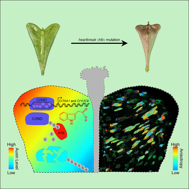
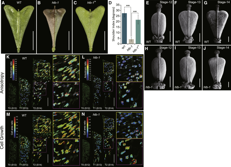
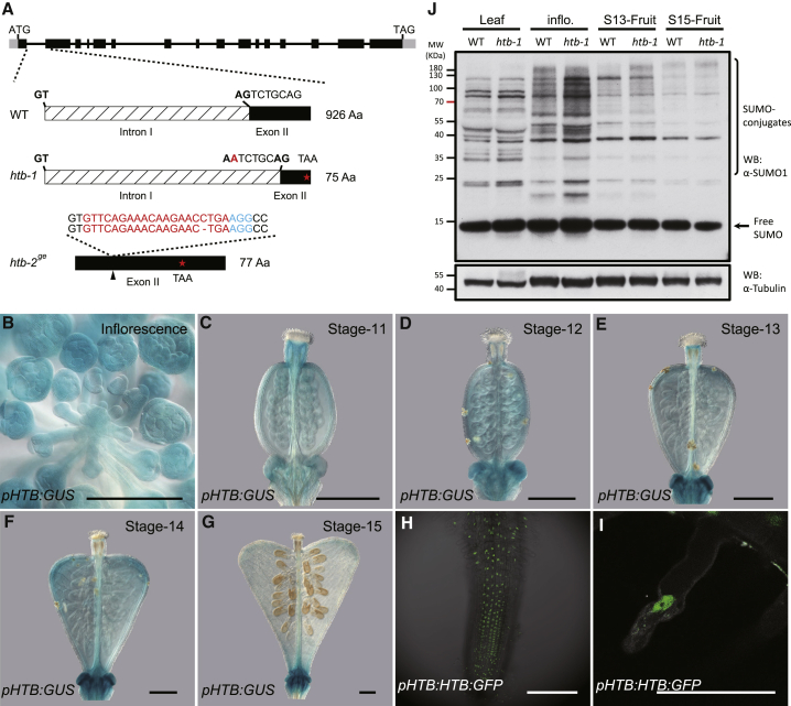
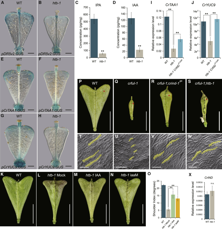
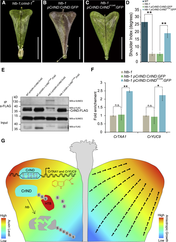
Morphological variation is the basis of natural diversity and adaptation. For example, angiosperms (flowering plants) evolved during the Cretaceous period more than 100 mya and quickly colonized terrestrial habitats [1]. A major reason for their astonishing success was the formation of fruits, which exist in a myriad of different shapes and sizes [2]. Evolution of organ shape is fueled by variation in expression patterns of regulatory genes causing changes in anisotropic cell expansion and division patterns [3-5]. However, the molecular mechanisms that alter the polarity of growth to generate novel shapes are largely unknown. The heart-shaped fruits produced by members of the Capsella genus comprise an anatomical novelty, making it particularly well suited for studies on morphological diversification [6-8]. Here, we show that post-translational modification of regulatory proteins provides a critical step in organ-shape formation. Our data reveal that the SUMO protease, HEARTBREAK (HTB), from Capsella rubella controls the activity of the key regulator of fruit development, INDEHISCENT (CrIND in C. rubella), via de-SUMOylation. This post-translational modification initiates a transduction pathway required to ensure precisely localized auxin biosynthesis, thereby facilitating anisotropic cell expansion to ultimately form the heart-shaped Capsella fruit. Therefore, although variation in the expression of key regulatory genes is known to be a primary driver in morphological evolution, our work demonstrates how other processes-such as post-translational modification of one such regulator-affects organ morphology.

Keywords: Capsella rubella; SUMOylation; anisotropic cell growth; fruit morphology; gene expression; post-translational modification.


Conflict of interest statement

Declaration of Interests The authors declare no competing interests.

Figures

None
Graphical abstract
Figure 1
Figure 1
The htb Mutant Produces Fruits with Defective Fruit Shape and Reduced Anisotropic Cell Growth (AC) Fruit morphology of WT (A), htb-1 (B), and rescue line of htb-1 transformed with pHTB:HTB:GFP (C) at stage 17. (D) Shoulder index measurements of fruits from WT, htb-1, and htb-1re (pHTB:HTB:GFP htb-1) plants. (E–G) Scanning electron microscopy (SEM) images of fruits from WT at developmental stages 12 (E), 13 (F), and 14 (G), showing fruit-shoulder growth after pollination. (H–J) SEM images of fruits from htb-1 at stages 12 (H), 13 (I), and 14 (J), showing compromised development of the fruit shoulders. (K–N) Time-lapse live imaging of developing fruits from stages 12 to 13 and 13 to 14 in WT (K and M) and htb-1 mutant (L and N). Cells are outlined by RFP signal of the clonal sectors derived from heat-shock treatment of pHS:CRE/BOB-lox line. The heatmaps represent the anisotropy (K and L) and the overall cell area ratio (M and N). Scale bars, (A–C) 5 mm; (E–N) 100 μm. Error bars in (D) represent SD of 30 individual fruits. ∗∗p < 0.01 (Student’s t test). See also Figure S1.
Figure 2
Figure 2
Molecular Cloning and Expression Analysis of HTB (A) Cloning of the htb-1 allele identified a G-to-A mutation in the acceptor site of the first intron of Carubv10008238, which disrupts the splicing of the first intron and results in a 7-bp deletion in the second exon, generating a premature stop codon in exon 2. The htb-2ge allele was generated by CRISPR with a single-base-pair deletion in the exon 2, resulting in a frameshift giving rise to a 77-amino-acid (aa) protein. The guide RNAs and PAM sequences were indicated by red and blue characters, respectively. (B–G) GUS staining of pHTB:GUS line showing the dynamic expression of HTB during fruit development. Uniform expression of HTB is detected in inflorescence tissue (B) and in the gynoecium at stage 11 (C) and 12 (D). A stronger HTB expression is detected in the developing fruit shoulders in stages 13 (E) and 14 (F). At stage 15, only residual HTB expression is observed in the fruit (G). (H and I) Subcellular localization of HTB:GFP protein in the roots of pHTB:HTB:GFP line. Scale bars in (B)–(I) represent 100 μm. (J) Comparative analysis of SUMO conjugates in total protein extracts from leaf, inflorescence (inflo.), and stage-13 (S13) and stage-15 (S15) fruits between WT and htb-1. The α-tubulin was immunoblotted as a loading control. See also Figure S2.
Figure 3
Figure 3
HTB Regulates Fruit Growth via Fine-Tuning Auxin Homeostasis (A and B) Auxin signaling visualized by pDR5v2:GUS in stage-14 fruits of WT (A) and htb-1 (B). (C and D) Measurements of IPA (C) and IAA (D) in fruit shoulders of WT and htb-1 in stage-14 fruits. (E–H) Expression of CrTAA1 and CrYUC9 shown by GUS staining of the pCrTAA1:GUS and pCrYUC9:GUS reporter lines at developmental stage 14 in WT (E and G) and htb-1 (F and H). (I and J) Expression analysis of CrTAA1 (I) and CrYUC9 (J) in fruit shoulders of WT, htb-1, and htb-1 pCrIND:CrINDK124R:GFP at stage 14. (K–N) Fruit morphology of WT (K), IAA mock (L), or IAA (M) treatment on htb-1 and htb-1 pCrIND:iaaM (N) at stage 17. (O) Shoulder index measurements of fruits from WT, htb-1 ± IAA treatment, and htb-1 pCrIND:iaaM plants. (P–S) Fruit morphology of WT (P), crful-1 (Q), crful-1;crind-1ge (R), and crful-1;htb-1 (S) at stage 17. Red dots indicate the location from where SEMs were taken in (T)–(W). (T–W) SEM images of valve epidermal cells of WT (T), crful-1 (U), crful-1;crind-1ge (V), and crful-1;htb-1 (W) at stage 17. (X) Expression analysis of CrIND in stage-14 fruits of WT and htb-1. n.s. indicates no statistically significant difference from WT. Scale bars in (A), (B), and (E)–(H), 150 μm; (K)–(N) and (P)–(S), 5 mm; and (T)–(W), 50 μM. Error bars in (C), (D), (I), (J), and (X) represent SD of three biological replicates and in (O) represent SD of 30 individual fruits. ∗∗p < 0.01 (Student’s t test). See also Figure S3 and Data S1 and S2.
Figure 4
Figure 4
HTB Stabilizes CrIND by De-SUMOylation (A–C) Fruit morphology of htb-1;crind-1ge (A), htb-1 pCrIND:CrIND:GFP (B), and htb-1 pCrIND:CrINDK124R:GFP (C) at stage 17. (D) Shoulder index measurements of fruits from WT, htb-1, htb-1 pCrIND:CrIND:GFP, and htb-1 pCrIND:CrINDK124R:GFP plants. (E) SUMOylation status of CrIND protein using pLhGR≫CrIND:FLAG and pLhGR≫CrINDK124R:FLAG lines. Immunoprecipitation experiments were conducted using anti-FLAG beads. Immunoblots were probed with anti-FLAG or anti-SUMO1 antibodies. (F) Chromatin immunoprecipitation (ChIP) analysis of CrIND/CrINDK124R associated with the CrYUC9 and CrTAA1 promoters. (G) Model for the control of heart-shape fruit development by CrIND and HTB in Capsella. Precise production of auxin in the tips of fruit shoulders controlled by CrIND induces anisotropic cell growth in the valves in a direction toward the shoulder tips. CrIND protein is de-SUMOylated by HTB, whereas in the htb-1 mutant, CrIND is SUMOylated and destabilized, thereby reducing its ability to activate expression of auxin biosynthesis gene (CrTAA1 and CrYUC9). Scale bars, (A–C) 5 mm. Error bars in (D) represent SD of 30 individual fruits and in (F) represent SD of three biological replicates. n.s. indicates no statistically significant difference from htb-1, *p < 0.05 and ∗∗p < 0.01 (Student’s t test). See also Figure S4.

References

    1. Soltis P.S., Folk R.A., Soltis D.E. Darwin review: angiosperm phylogeny and evolutionary radiations. Proc. Royal Soc. B: Biol. Sci. 2019;286:20190099.
    1. Seymour G.B., Østergaard L., Chapman N.H., Knapp S., Martin C. Fruit development and ripening. Annu. Rev. Plant Biol. 2013;64:219–241. - PubMed
    1. Carroll S.B. Endless forms: the evolution of gene regulation and morphological diversity. Cell. 2000;101:577–580. - PubMed
    1. Carroll S.B. Evolution at two levels: on genes and form. PLoS Biol. 2005;3:e245. - PMC - PubMed
    1. Wray G.A. The evolutionary significance of cis-regulatory mutations. Nat. Rev. Genet. 2007;8:206–216. - PubMed

Publication types

MeSH terms

LinkOut - more resources