. 2020 Sep 15;95(11):e1512-e1527.
doi: 10.1212/WNL.0000000000010327. Epub 2020 Aug 13.

The clinical, histologic, and genotypic spectrum of SEPN1-related myopathy: A case series

Affiliations

The clinical, histologic, and genotypic spectrum of SEPN1-related myopathy: A case series

Rocio N Villar-Quiles et al. Neurology. .

Abstract

Objective: To clarify the prevalence, long-term natural history, and severity determinants of SEPN1-related myopathy (SEPN1-RM), we analyzed a large international case series.

Methods: Retrospective clinical, histologic, and genetic analysis of 132 pediatric and adult patients (2-58 years) followed up for several decades.

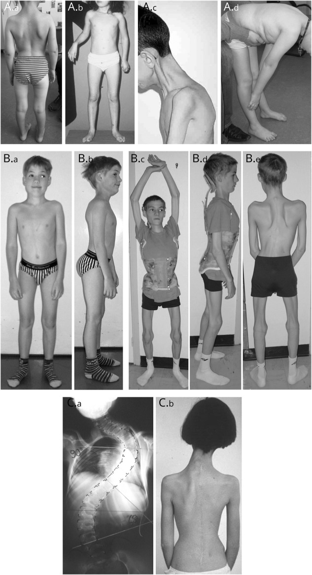
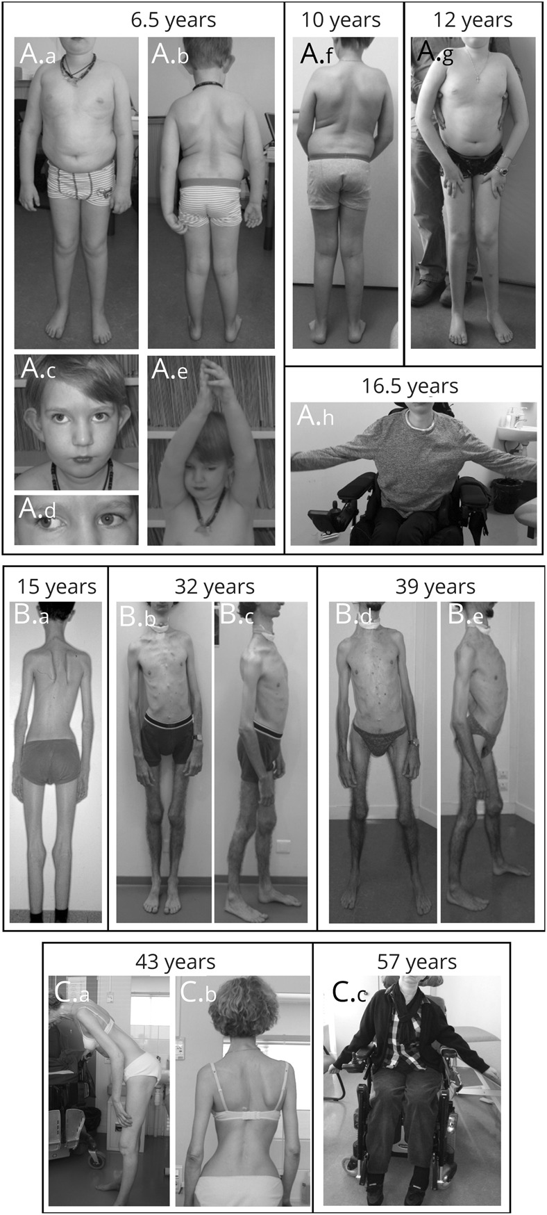
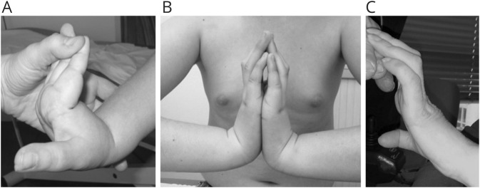
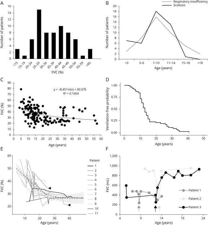
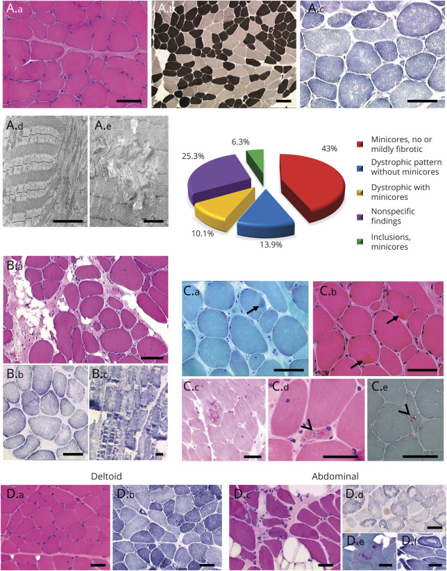
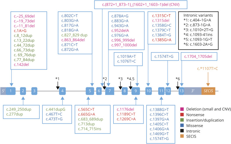
Results: The clinical phenotype was marked by severe axial muscle weakness, spinal rigidity, and scoliosis (86.1%, from 8.9 ± 4 years), with relatively preserved limb strength and previously unreported ophthalmoparesis in severe cases. All patients developed respiratory failure (from 10.1±6 years), 81.7% requiring ventilation while ambulant. Histopathologically, 79 muscle biopsies showed large variability, partly determined by site of biopsy and age. Multi-minicores were the most common lesion (59.5%), often associated with mild dystrophic features and occasionally with eosinophilic inclusions. Identification of 65 SEPN1 mutations, including 32 novel ones and the first pathogenic copy number variation, unveiled exon 1 as the main mutational hotspot and revealed the first genotype-phenotype correlations, bi-allelic null mutations being significantly associated with disease severity (p = 0.017). SEPN1-RM was more severe and progressive than previously thought, leading to loss of ambulation in 10% of cases, systematic functional decline from the end of the third decade, and reduced lifespan even in mild cases. The main prognosis determinants were scoliosis/respiratory management, SEPN1 mutations, and body mass abnormalities, which correlated with disease severity. We propose a set of severity criteria, provide quantitative data for outcome identification, and establish a need for age stratification.

Conclusion: Our results inform clinical practice, improving diagnosis and management, and represent a major breakthrough for clinical trial readiness in this not so rare disease.


Figures

Figure 1
Figure 1. Distinctive clinical signs
(A) The clinical phenotype in early childhood was recognizable by predominantly axial weakness, deltoid and inner thigh amyotrophy (“bracket-like thighs”) (A.a and A.b) and spinal rigidity identifiable upon specific examination (A.c and A.d). Axial rigidity was typically more prominent in the cervicodorsal spine (A.c and A.d), with relative or full preservation of lumbar spine flexion (A.d). (B) Typical moderate patient at ages 11 (B.a and B.b) and 14 years (B.c–B.e). Often subtle until late childhood, the full phenotype usually became apparent around puberty. Body weight decreased dramatically, leading in some cases to an apparent loss of subcutaneous adipose tissue (lower limbs in B.c and B.d) and a cachexia phenotype. Most patients developed scoliosis (B.e), which required adapted bracing to avoid thorax compression (Garchois brace, B.c and B.d). SEPN1-RM scoliosis is recognizable due to dorsal lordosis leading to pseudo-scapular winging, lateral trunk deviation with compensatory contralateral neck shift, and horizontally aligned hips. (C) Scoliosis progression despite bracing often required arthrodesis (moderately affected patient before [C.a] and after [C.b] spinal fusion).
Figure 2
Figure 2. Disease course in typical severe, moderate, and mild cases
(A) This severely affected patient presented with neonatal axial muscle weakness, required nighttime assisted ventilation since diagnosis at 6.5 years of age, and showed at that age sternocleidomastoid, deltoid, and leg amyotrophy (A.a and A.b), antigravity limb strength (A.e), and ophthalmoparesis with limited upward (A.c.) and horizontal (A.d.) eye movements. Note the typical SEPN1-related myopathy (SEPN1-RM) facial appearance (tubular nose, prominent nasal sella, low-set prominent ears and mild retrognathia, midsegment hypotrophy [A.c]), and increased trunk adiposity (A.a and A.b). He developed scoliosis from age 7 years (A.f), which eventually required arthrodesis, lost independent ambulation at 11 years (A.g), and was wheelchair-bound with severe limb weakness at 16 years (A.h, maximum antigravity power of upper limbs). (B) After spinal fusion and instauration of nocturnal ventilation through a tracheostomy at 15 years of age (B.a), this patient with moderate SEPN1-RM remained stable until the middle of the fourth decade (B.b and B.c), when progressive limb weakness led to a wider base of support, increased hyperlordosis (B.d and B.e), and limited upper limb abduction and ambulation. Note reduced anteroposterior thorax diameter, severe diffuse amyotrophy, and loss of subcutaneous fatty tissue. (C) One of the mildest cases in this series resulted in normal motor development including head control, rigid spine (C.a), and mild untreated scoliosis (C.b) and required assisted ventilation only after the age of 35 years. A myopathy was first suspected in the fourth decade due to progression of a previously mild muscle weakness. Continuous progression eventually led to loss of antigravity movements and ambulation and almost permanent ventilation by the age of 57 years (C.c, maximum upper limb abduction) and to death at age 58 years.
Figure 3
Figure 3. Hand hyperlaxity and finger flexor contractures
Moderate distal hyperlaxity was common in early childhood (A, 7-year-old patient) and often coexisted with finger flexor contractures visible upon wrist extension. The latter tended to appear from adolescence (B, 16-year-old patient) and became more prominent with age (C, 57-year-old patient).
Figure 4
Figure 4. Respiratory involvement
(A) Distribution of forced vital capacity (FVC) (% of predicted values) in 69 cases shows that most patients had FVC <39%, the most frequent range being between 25% and 29%. (B) Respiratory insufficiency (gray line) or scoliosis (black line) were first detected in most patients around the same age, most commonly between 7 and 10 years. (C) Progression of FVC in 32 patients (ages 4–58 years) revealed decrease of FVC with age in years using a logarithmic regression (R2 = 0.16642). Individual values are shown as black dots. (D) Kaplan-Meier curve showing ventilation-free probability with age. (E) Progression of respiratory involvement in 11 patients, revealing extreme variability between patients with similar disease severity. Note also intrapatient variability in FVC values (arrowheads). (F) Follow-up of respiratory involvement before and after surgical correction of scoliosis in 3 patients. FVC typically dropped in the postsurgical period (arrows) and then came back to previous or even higher values with correct postoperative management including ventilation.
Figure 5
Figure 5. Histopathologic patterns and different histologic severity in axial vs limb muscles
The relative frequency of the different histopathologic patterns observed in this series (n = 78) is represented in the graph. Foci of sarcomere disorganization and mitochondria depletion, spanning only a few sarcomeres on the longitudinal axis of the fiber (minicores), were observed in 60% of the biopsies (A.c–A.e and B.b and B.c). (A) The most common pattern was typical of a congenital myopathy with minicores (multi-minicore disease), including mild or no endomysial fibrosis (A.a), type I fiber predominance and relative hypotrophy (dark fibers, A.b), and multiple lighter zones devoid of succinate dehydrogenase (SDH) or nicotinamide adenine dinucleotide (NADH) enzymatic activity (A.c) corresponding to mitochondria depletion and sarcomere disorganization on electron microscopy (EM) (minicores) (A.d and A.e). (B) Around 24% of biopsies showed a mild to moderate congenital muscular dystrophy pattern, associated with either abundant or scattered/inconspicuous minicores. Note prominent endomysial fibroadiposis but rare necrotic or regenerating fibers (B.a). (C) Eosinophilic inclusions compatible with Mallory body–like inclusions (arrows, C.a and C.b) or rimmed vacuoles (arrowheads, C.d and C.e) were identified in some samples but typically involved a small percentage of fibers. Thus they were easily overlooked unless specifically searched for. (D) Two muscle samples taken from the same patient at 12 years of age revealed that histopathologic changes were more severe in axial than in limb muscles, mirroring the clinical situation. Her deltoid muscle (D.a and D.b) showed minor myopathic changes, with mild fiber size variation and scattered internalized nuclei and minicores. In contrast, her abdominal muscles revealed major dystrophic changes with muscle fiber loss, fatty-adipose replacement, rimmed vacuoles (D.c), rod-like inclusions (D.e), multi-minicores (D.d), and also well-delimited cores with a hyperoxidative perilesional rim (similar to those observed in central core disease) (D.f). Transversal frozen sections stained with hematoxylin & eosin (A.a, B.a, C.b–C.d, D.a and D.c), reduced NADH (A.c, B.c, D.b and D.f), ATPase 4.6 (A.b), SDH (B.b and D.d), modified Gomori trichrome (C.a and C.e, D.e); longitudinal EM sections (A.d and A.e). Scale bars: 50 µm except for A.d and A.e: 2 µm.
Figure 6
Figure 6. Schematic representation of the SEPN1 (SELENON) gene and localization of the identified mutations
Exons are depicted in light blue and the 3′ untranslated region (UTR) SECIS element in orange. The putative reductase domain containing the only selenocysteine residue in SEPN1 is encoded by exon 10. The EF-hand domain, potentially calcium-binding, is encoded by exon 2. Color code for variants: pink, deletion; green, insertions/duplications; blue, missense; red, nonsense; black, intronic variants affecting splicing; orange, variants affecting SEPN1 3′ UTR SECIS element. CNV = copy number variation; SECIS = selenocysteine insertion sequence.

References

    1. Moghadaszadeh B, Desguerre I, Topaloglu H, et al. . Identification of a new locus for a peculiar form of congenital muscular dystrophy with early rigidity of the spine, on chromosome 1p35-36. Am J Hum Genet 1998;62:1439–1445. - PMC - PubMed
    1. Lescure A, Gautheret D, Carbon P, Krol A. Novel selenoproteins identified in silico and in vivo by using a conserved RNA structural motif. J Biol Chem 1999;274:38147–38154. - PubMed
    1. Moghadaszadeh B, Petit N, Jaillard C, et al. . Mutations in SEPN1 cause congenital muscular dystrophy with spinal rigidity and restrictive respiratory syndrome. Nat Genet 2001;29:17–18. - PubMed
    1. Ferreiro A, Quijano-Roy S, Pichereau C, et al. . Mutations of the selenoprotein N gene, which is implicated in rigid spine muscular dystrophy, cause the classical phenotype of multiminicore disease: reassessing the nosology of early-onset myopathies. Am J Hum Genet 2002;71:739–749. - PMC - PubMed
    1. Clarke NF, Kidson W, Quijano-Roy S, et al. . SEPN1: associated with congenital fiber-type disproportion and insulin resistance. Ann Neurol 2006;59:546–552. - PubMed

Publication types