Review
. 2020 Aug 11;9(8):2596.
doi: 10.3390/jcm9082596.

Mechanisms of Mitochondrial Dysfunction in Lysosomal Storage Disorders: A Review

Affiliations
Review

Mechanisms of Mitochondrial Dysfunction in Lysosomal Storage Disorders: A Review

Karolina M Stepien et al. J Clin Med. .

Abstract

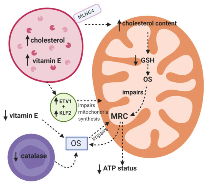
Mitochondrial dysfunction is emerging as an important contributory factor to the pathophysiology of lysosomal storage disorders (LSDs). The cause of mitochondrial dysfunction in LSDs appears to be multifactorial, although impaired mitophagy and oxidative stress appear to be common inhibitory mechanisms shared amongst these heterogeneous disorders. Once impaired, dysfunctional mitochondria may impact upon the function of the lysosome by the generation of reactive oxygen species as well as depriving the lysosome of ATP which is required by the V-ATPase proton pump to maintain the acidity of the lumen. Given the reported evidence of mitochondrial dysfunction in LSDs together with the important symbiotic relationship between these two organelles, therapeutic strategies targeting both lysosome and mitochondrial dysfunction may be an important consideration in the treatment of LSDs. In this review we examine the putative mechanisms that may be responsible for mitochondrial dysfunction in reported LSDs which will be supplemented with morphological and clinical information.

Keywords: autophagy; inflammation; lysosomal storage diseases; mitochondrial dysfunction; mitophagy and cytokine; oxidative stress; reactive oxygen species.


Conflict of interest statement

The authors declare no conflict of interest.

Figures

Figure 1
Figure 1
Putative mechanisms responsible for mitochondrial dysfunction in Niemann Pick C. MRC: Mitochondrial respiratory chain. ETV1: Transcription factor. KLF2: Transcription factor. OS: Oxidative stress. GSH: Reduced glutathione.
Figure 2
Figure 2
The putative mechanisms that have been implicated in inducing mitochondrial dysfunction in Pompe disease. MRC: Mitochondrial respiratory chain. Ca2+: Calcium. MTORC1: mammalian target of rapamycin complex 1. ROS: Reactive oxygen species.
Figure 3
Figure 3
The muscle biopsy from a patient with late-onset Pompe disease (PD) shows myopathic changes including atrophic and hypertrophic fibers; one fiber with subsarcolemmal accumulation of mitochondria is also present (asterisk). (A)—hematoxylin-eosin, ×20; staining for acid phosphatase highlights the increase in lysosomes in muscle fibers. (B)—acid phosphatase, ×20; several COX-negative fibers (blue) are documented in the combined staining COX/SDH. (C)—COX/SDH, ×20.
Figure 4
Figure 4
Putative mechanisms responsible for mitochondrial dysfunction in Mucopolysaccharidosis. HS: Heparin sulphate. MRC: Mitochondrial respiratory chain. ROS: Reactive oxygen species. RNS: Reactive nitrogen species. TNF: Cytokine.
Figure 5
Figure 5
These images document the muscle from a patient with Fabry’s disease on ERT treatment; the biopsy is characterized by abnormal variation in fibre size and a few fibres showing internal nucleation (A, hematoxylin-eosin, ×20); focal subsarcolemmal accumulation of mitochondria but no ragged red fibres is seen (arrow) (B—modified Gomori’s trichrome, ×40); a COX-negative fibre is present (blue fibre, arrow) (C—COX/SDH staining, ×40); there is increase staining with acid phosphatase (red precipitate) that due to subsarcolemmal deposits of lipofuscins and increase in lysosomes (D—acid phosphatase, ×20).
Figure 6
Figure 6
Putative causes of mitochondrial dysfunction in Fabry disease. GB3: globotriaosylceramide. Lacg: Lacoglyceramide. α-galA: alpha-galactosidase A. SOD-2: Superoxide dismutase-2. eNOS:endothelial nitric oxide synthase. iNOS: inducible nitric oxide synthase. MRC: Mitochondrial respiratory chain. GSH: Reduced glutathione. ROS: Reactive oxygen species. RNS: Reactive oxygen species. NO: Nitric oxide.

References

    1. Platt F.M., Boland B., van der Spoel A.C. Lysosomal storage disorders: The cellular impact of lysosomal dysfunction. J. Cell Biol. 2012;199:723–734. doi: 10.1083/jcb.201208152. - DOI - PMC - PubMed
    1. Parkinson-Lawrence E.J., Shandala T., Prodoehl M., Plew R., Borlace G.N., Brooks D.A. Lysosomal storage disease: Revealing lysosomal function and physiology. Physiology. 2010;25:102–115. doi: 10.1152/physiol.00041.2009. - DOI - PubMed
    1. Echevarria L., Benistan K., Toussaint A., Dubourg O., Hagege A.A., Eladari D., Jabbour F., Beldjord C., De Mazancourt P., Germain D.P. X-chromosome inactivation in female patients with Fabry disease. Clin. Genet. 2016;89:44–54. doi: 10.1111/cge.12613. - DOI - PubMed
    1. Wraith J.E. Lysosomal disorders. Semin. Neonatol. 2002;7:75–83. doi: 10.1053/siny.2001.0088. - DOI - PubMed
    1. Audano M., Schneider A., Mitro N. Mitochondria, lysosomes, and dysfunction: Their meaning in neurodegeneration. J. Neurochem. 2018;147:291–309. doi: 10.1111/jnc.14471. - DOI - PubMed

LinkOut - more resources