. 2020 Aug 11;21(16):5763.
doi: 10.3390/ijms21165763.

Vascular Remodeling in Moyamoya Angiopathy: From Peripheral Blood Mononuclear Cells to Endothelial Cells

Affiliations

Vascular Remodeling in Moyamoya Angiopathy: From Peripheral Blood Mononuclear Cells to Endothelial Cells

Francesca Tinelli et al. Int J Mol Sci. .

Abstract

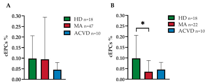
The pathophysiological mechanisms of Moyamoya angiopathy (MA), which is a rare cerebrovascular condition characterized by recurrent ischemic/hemorrhagic strokes, are still largely unknown. An imbalance of vasculogenic/angiogenic mechanisms has been proposed as one possible disease aspect. Circulating endothelial progenitor cells (cEPCs) have been hypothesized to contribute to vascular remodeling of MA, but it remains unclear whether they might be considered a disease effect or have a role in disease pathogenesis. The aim of the present study was to provide a morphological, phenotypical, and functional characterization of the cEPCs from MA patients to uncover their role in the disease pathophysiology. cEPCs were identified from whole blood as CD45dimCD34+CD133+ mononuclear cells. Morphological, biochemical, and functional assays were performed to characterize cEPCs. A significant reduced level of cEPCs was found in blood samples collected from a homogeneous group of adult (mean age 46.86 ± 11.7; 86.36% females), Caucasian, non-operated MA patients with respect to healthy donors (HD; p = 0.032). Since no difference in cEPC characteristics and functionality was observed between MA patients and HD, a defective recruitment mechanism could be involved in the disease pathophysiology. Collectively, our results suggest that cEPC level more than endothelial progenitor cell (EPC) functionality seems to be a potential marker of MA. The validation of our results on a larger population and the correlation with clinical data as well as the use of more complex cellular model could help our understanding of EPC role in MA pathophysiology.

Keywords: Moyamoya angiopathy; RNF213; endothelial progenitor cells; neovascularization.


Conflict of interest statement

The authors declare no conflict of interest. The funders had no role in the design of the study; in the collection, analyses, or interpretation of data; in the writing of the manuscript, or in the decision to publish the results.

Figures

Figure 1
Figure 1
Endothelial progenitor cell (EPC) level in whole blood (WB) from (A) a heterogeneous and (B) a homogeneous (adult, Caucasian, non-operated) group of Moyamoya angiopathy (MA) patients, as compared with healthy donors (HD) and atherosclerotic cerebrovascular disease (ACVD) controls: Data are expressed as mean of circulating endothelial progenitor cell (cEPC)% ± standard deviation (SD), where cEPC% was calculated as follows: (cEPCs/µL/WB cells/μL) × 100; statistical significance (* p < 0.05) was calculated through Student’s t-test (p = 0.032).
Figure 2
Figure 2
Cultured EPCs from (AE) HD, (FJ) MA, and (KO) ACVD representative subjects at 5, 7, 10, 17, and 31 days after seeding in Microvascular Endothelial Cell Growth Medium (EGM-MV medium) (20× magnification, 50 μm scale bar).
Figure 3
Figure 3
Staining of late EPCs obtained from a representative MA patient by (A) Bright field and (BD) fluorescence microscopy; (B) DAPI, (C) FITC-Ulex-lectin, (D) DAPI+FITC-Ulex-lectin (20× magnification, 50 μm scale bar).
Figure 4
Figure 4
mRNA relative expression of (A) von Willebrand Factor (vWF), (B) CD31, and (C) KDR/ Vascular endothelial growth factor-receptor2 (VEGF-R2) in EPCs at 17 and 31 days after seeding: the mRNA levels in EPCs at day 31 from HD, MA, and ACVD subjects are expressed in relation to EPCs at day 17, arbitrarily imposed at 1 as calibrator. β2-microglobulin (β2M) was used as housekeeping gene. Data were expressed as mean ± SD, and statistical significance (* p < 0.05, ** p < 0.01) was calculated through Student’s t-test. Values of at least three independent experiments are shown.
Figure 5
Figure 5
Tube formation assay on Human Umbilical Vein Endothelial Cell (HUVEC) cells in the presence of conditioned media from HD and MA EPC cultures at (A,C) day 7 and (B,D) day 17 (representative images are shown; 5× magnification, 12.5 μm scale bar): The angiogenic potential of the growth factors released by early and late EPCs was evaluated through the analysis of the photos from each condition by the Wimasis software. Three independent experiments for each condition have been considered.
Figure 6
Figure 6
Vasculogenic capacity of HUVEC cells cultured in conditioned media collected (A) 7 and (B) 17 days after seeding of EPCs from HD and from a heterogeneous group of MA patients (each different tube parameter has been normalized with values obtained by HUVEC cells in EGM-MV medium): Data were expressed as mean ± SD, and statistical significance was calculated through Student’s t-test. Values of at least three independent experiments are shown.
Figure 7
Figure 7
Vasculogenic capacity of HUVEC cells cultured in conditioned media collected (A) 7 and (B) 17 days after seeding of EPCs from HD and from a homogeneous group of MA patients (each different tube parameter has been normalized with values obtained by HUVEC cells in EGM-MV medium): Data were expressed as mean ± SD, and statistical significance was calculated through Student’s t-test. Values of at least three independent experiments are shown.
Figure 8
Figure 8
(A) Vascular endothelial growth factor A (VEGF-A), (B) hepatocyte growth factor (HGF), (C) transforming growth factor-beta 1 (TGF-β1), (D) chemokine (C-C motif) ligand 5 (CCL5/RANTES), (E) chemokine (C-C motif) ligand 2 (CCL2/MCP-1), and (F) interleukin 8 (IL-8/CXCL8) concentration (pg/mL) in conditioned media collected from EPC cultures at 7 and 17 days after seeding: Data were expressed as mean ± SD, and statistical significance (* p < 0.05) was calculated through Student’s t-test. Values of at least three independent experiments are shown.
Figure 9
Figure 9
mRNA relative expression of CD31, HGF, and TGF-β1 in EPCs at 7 days after seeding: The mRNA levels in EPC from MA patients are expressed in relation to HD subjects, arbitrarily imposed at 1 as calibrator. β2M was used as housekeeping gene. Data were expressed as mean ± SD, and statistical significance (* p < 0.05) was calculated through Student’s t-test. Values of at least three independent experiments are shown.

References

    1. Fukui M. Guidelines for the diagnosis and treatment of spontaneous occlusion of the circle of Willis (’moyamoya’ disease). Research committee on spontaneous occlusion of the circle of willis (moyamoya disease) of the ministry of health and welfare, Japan. Clin. Neurol. Neurosurg. 1997;99:S238–S240. doi: 10.1016/S0303-8467(97)00082-6. - DOI - PubMed
    1. Bersano A., Guey S., Bedini G., Nava S., Hervé D., Vajkoczy P., Tatlisumak T., Sareela M., Van Der Zwan A., Klijn C.J., et al. Research progresses in understanding the pathophysiology of moyamoya disease. Cerebrovasc. Dis. 2016;41:105–118. doi: 10.1159/000442298. - DOI - PubMed
    1. Kossorotoff M., Tournier-Lasserve E., Herve D., Guey S. Moyamoya disease and syndromes: From genetics to clinical management. Appl. Clin. Genet. 2015;8:49–68. doi: 10.2147/TACG.S42772. - DOI - PMC - PubMed
    1. Kobayashi E., Saeki N., Oishi H., Hirai S., Yamaura A. Long-term natural history of hemorrhagic type moyamoya disease in 42 patients. J. Neurosurg. 2000;93:976–980. doi: 10.3171/jns.2000.93.6.0976. - DOI - PubMed
    1. Kraemer M., Heienbrok W., Berlit P. Moyamoya Disease in Europeans. Stroke. 2008;39:3193–3200. doi: 10.1161/STROKEAHA.107.513408. - DOI - PubMed

MeSH terms