. 2020 Aug 14;5(1):156.
doi: 10.1038/s41392-020-00263-y.

Adaptive immune responses to SARS-CoV-2 infection in severe versus mild individuals

Affiliations

Adaptive immune responses to SARS-CoV-2 infection in severe versus mild individuals

Fan Zhang et al. Signal Transduct Target Ther. .

Erratum in

Abstract

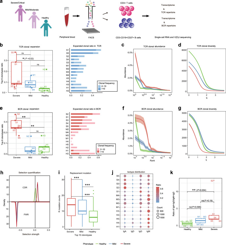
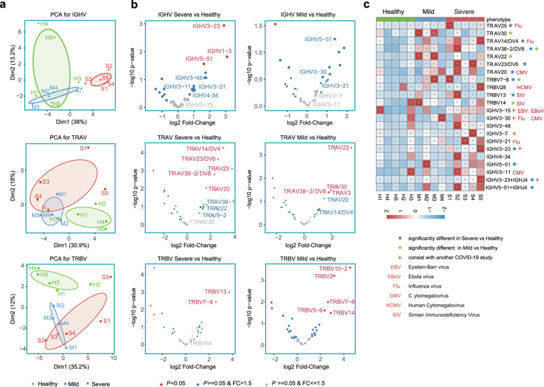
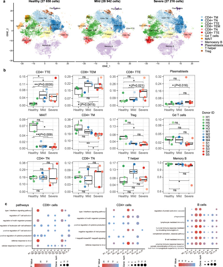
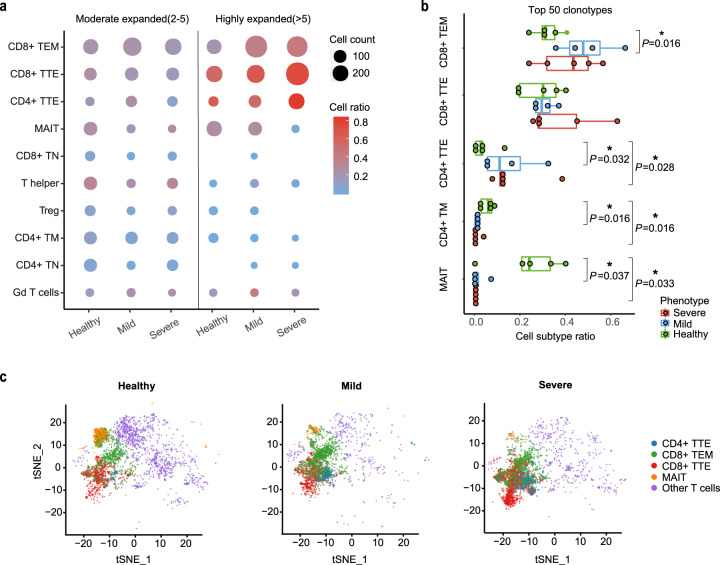
The global Coronavirus disease 2019 (COVID-19) pandemic caused by SARS-CoV-2 has affected more than eight million people. There is an urgent need to investigate how the adaptive immunity is established in COVID-19 patients. In this study, we profiled adaptive immune cells of PBMCs from recovered COVID-19 patients with varying disease severity using single-cell RNA and TCR/BCR V(D)J sequencing. The sequencing data revealed SARS-CoV-2-specific shuffling of adaptive immune repertories and COVID-19-induced remodeling of peripheral lymphocytes. Characterization of variations in the peripheral T and B cells from the COVID-19 patients revealed a positive correlation of humoral immune response and T-cell immune memory with disease severity. Sequencing and functional data revealed SARS-CoV-2-specific T-cell immune memory in the convalescent COVID-19 patients. Furthermore, we also identified novel antigens that are responsive in the convalescent patients. Altogether, our study reveals adaptive immune repertories underlying pathogenesis and recovery in severe versus mild COVID-19 patients, providing valuable information for potential vaccine and therapeutic development against SARS-CoV-2 infection.


Conflict of interest statement

The authors declare no competing interests.

Figures

Fig. 1
Fig. 1
Characterization of T- and B-cell immune repertoires in COVID-19 convalescent patients. a Schematic representation of overall study design. Blood samples from recovered patients with severe/critical infection or mild/moderate infection, and healthy controls were collected and sorted to obtain CD3+ T cells and CD19+CD20+CD27+ antigen-experienced B cells (AEBCs). Sorted cells underwent high-throughput single-cell RNA and TCR/BCR V(D)J sequencing for further analyses. Characterization and comparison of TCR clonal expansion among severe/critical patients (SPs, red), mild/moderate patients (MPs, blue), and healthy controls (HCs, green), by quantifying the ratio of expanded clones (b), clonal abundance (c), and clonal diversity (d). Characterization and comparison of BCR clonal expansion among SPs, MPs, and HCs, by quantifying the ratio of expanded clones (e), clonal abundance (f), and clonal diversity (g). h Comparison of selection strength on complementary-determining region (CDR, top) and framework region (FWR, bottom) of the BCRs among SPs, MPs, and HCs by density plot. i Comparison of replacement mutation frequency on IGHV gene segments among SPs, MPs, and HCs by boxplot. j Comparison of isotype distributions among SPs, MPs, and HCs. k Comparison of the ratio of (IgA + IgG) to (IgD + IgM) in SPs, MPs, and HCs by boxplot. ***P < 0.001; **P < 0.01; *P < 0.05; n.s. not significant using Wilcoxon rank sum test for pairwise comparisons
Fig. 2
Fig. 2
Comparison of V gene usage of IGH, TRA, and TRB among SPs, MPs, and HCs. a Principal component analysis (PCA) for IGHV-gene usage and TRAV or TRBV-gene usage in SPs (S1–S5), MPs (M1–M4), and HCs (H1–H5). b Volcano plot representing differences of IGHV-gene usage in BCR repertoires or TRAV/TRBV-gene usage of TCR repertoires. Positive fold-change values denote more frequent IGHV genes or TRAV/TRBV genes in COVID-19 patients. Genes with P value < 0.05 are displayed in red. Genes with a P value > 0.05 but fold-change (FC) value > 1.5 are displayed in blue. c Heatmap of reprehensive gene segments enriched in SPs or MPs compared with in HCs. Colors denote frequencies of each V gene segment used in each sample. The V genes overpresented in SPs or MPs (P value < 0.05) are indicated using a blue or a green star, respectively. Other known biasedly used gene segments related to virus-specific antibodies are marked using relevant virus name (EBV, EBoV, Flu, CMV, HCMV, and SIV). Genes consistent with another COVID-19 recovered patient study are denoted with a cross
Fig. 3
Fig. 3
Characterization of variations in cell composition and functional status of the peripheral T and B cells of the recovered COVID-19 patients. a Two-dimensional t-SNE visualization of 12 major cell types identified from healthy controls (HCs, left), mild patients (MPs, middle), and severe patients (SPs, right) including four for CD8+ T cells, five for CD4+ T cells, one for γδ T cells, and two for B cells. Each dot corresponds to one single cell, and colored according to cell types. The number of total cells showed in each t-SNE map was labeled on the top. b Boxplot representing proportions of each cell type in each sample. Each dot corresponds to one sample, and colored by group-specific color scheme (severe, red gradients; mild, blue gradients; healthy, green gradients). **P < 0.01; *P < 0.05; n.s. not significant by Wilcoxon rank sum test. c Dot plot depicting upregulated canonical pathways in CD4+ T cells, CD8+ T cells, and AEBCs shared by multiple COVID-19 patients. Differentially expressed genes and their enriched pathways were calculated as each COVID-19 patient relative to all healthy controls (S# versus H or M# versus H), and each severe patient relative to all mild cases (S# versus M)
Fig. 4
Fig. 4
Comparison of TCR clonal expansion in T-cell subsets among severe patients (SPs), mild patients (MPs) and healthy controls (HCs). a Dot plot depicting clonal expansion status of distinct T-cell subsets in HCs, MPs, and SPs. Dot size denotes cell counts in each T-cell subset belonging to more expanded (frequency > 5) or less expanded (2 ≤ frequency ≤ 5) clonotypes. Red-blue color range denotes the ratio of expanded cells occupied in each T-cell subset. b Boxplot depicting the top 50 TCR clonotypes in each donor consists of various T-cell subsets of different proportions. *P < 0.05; n.s. not significant using Wilcoxon rank sum test for pairwise comparisons. c T-SNE projection of clonally expanded T cells (blue, CD4+ TTE; green, CD8+ TEM; red, CD8+ TTE; orange, MAIT; violet, other T cells) in HCs, MPs, and SPs
Fig. 5
Fig. 5
Assessment of SARS-CoV-2 specific T-cell immunity in COVID-19 convalescent patients by IFN-γ ELISpot and FACS analysis. a Composition and fraction of peptides used in ten SARS-CoV-2 antigen pools. b Boxplot comparing the number of spot-forming cells (y axis) from ELISpot assay between stimulated severe and mild samples for each antigen group (x axis). Each donor was tested in duplicates. For each donor, the average number of spots from stimulated samples subtract that of the unstimulated samples was shown as a dot on the plot. Negative values were set to zero. Red and blue dots indicate SPs and MPs, respectively. ***P < 0.001 by unpaired Student’s t test. SFC spot-forming cells. The blue or red text on top indicates the percentage of stimulated mild or severe donors shown positive in ELISPOT, respectively. Antigen-specific CD69+CD8+ T cell (c) and CD4+ T cell (d) reactivity in ELISpot-positive COVID-19 cases between the negative control (DMSO, blue dots) and antigen pool stimulations (red dots). **P < 0.01; *P < 0.05 by paired t-test. e Pearson’s correlation between SARS-CoV-2-specific CD4+ T cells (x axis) and SARS-CoV-2-specific CD8+ T cells (y axis)

References

    1. Long, Q. X. et al. Antibody responses to SARS-CoV-2 in patients with COVID-19. Nat. Med.10.1038/s41591-020-0897-1 (2020). - PubMed
    1. Yu F, Du L, Ojcius DM, Pan C, Jiang S. Measures for diagnosing and treating infections by a novel coronavirus responsible for a pneumonia outbreak originating in Wuhan, China. Microbes Infect. 2020;22:74–79. doi: 10.1016/j.micinf.2020.01.003. - DOI - PMC - PubMed
    1. Liao, M. et al. Single-cell landscape of bronchoalveolar immune cells in patients with COVID-19. Nat. Med. 10.1038/s41591-020-0901-9 (2020). - PubMed
    1. Wilk, A. J. et al. A single-cell atlas of the peripheral immune response to severe COVID-19. 10.1101/2020.04.17.20069930 (2020). - PMC - PubMed
    1. Li T, et al. Significant changes of peripheral T lymphocyte subsets in patients with severe acute respiratory syndrome. J. Infect. Dis. 2004;189:648–651. doi: 10.1086/381535. - DOI - PMC - PubMed

Publication types

MeSH terms