. 2020 Aug 14;11(8):624.
doi: 10.1038/s41419-020-02849-4.

Ischemia induces autophagy of endothelial cells and stimulates angiogenic effects in a hindlimb ischemia mouse model

Affiliations

Ischemia induces autophagy of endothelial cells and stimulates angiogenic effects in a hindlimb ischemia mouse model

In-Hye Jeong et al. Cell Death Dis. .

Abstract

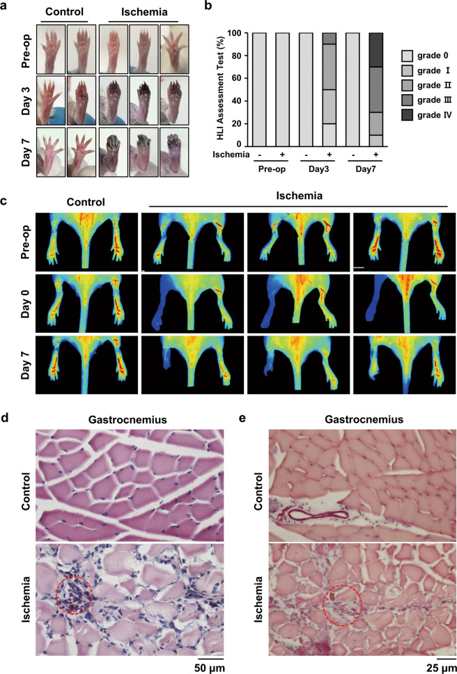
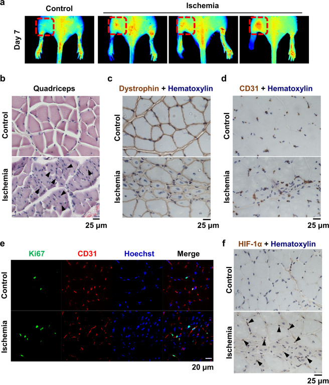
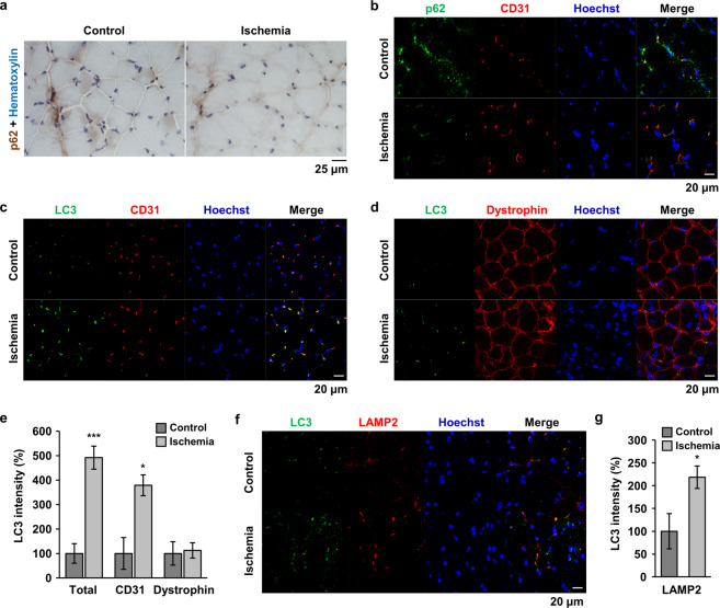
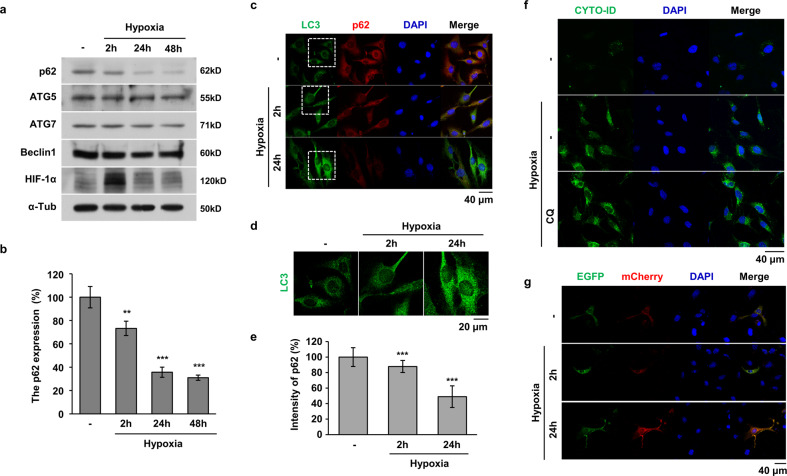
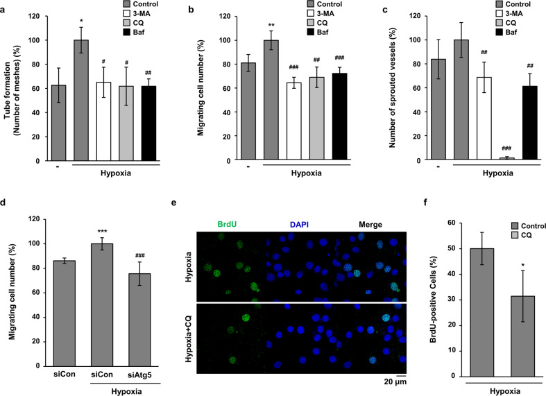
Although peripheral artery disease (PAD) is a major health problem, there have been limited advances in medical therapies. In PAD patients, angiogenesis is regarded as a promising therapeutic strategy to promote new arterial vessels and improve perfusion of ischemic tissue. Autophagy plays a critical role in catabolic processes for cell survival under normal and stressful conditions and plays fundamental biological roles in various cellular functions. In the present study, we showed that autophagy in endothelial cells is important for the repair and regeneration of damaged tissues. In a hindlimb ischemia mouse model, autophagy was stimulated in endothelial cells of the quadriceps muscle, and adjacent cells proliferated and regenerated. The autophagy pathway was induced under prolonged hypoxia in endothelial cells, and autophagy increased angiogenic activities. Moreover, conditioned media from endothelial cells blocked autophagy and inhibited the proliferation of muscle cells, suggesting that autophagic stimulation in endothelial cells affects the survival of adjacent cells, such as muscle. Collectively, hypoxia/ischemia-induced autophagy angiogenesis, and the damaged tissue surrounded by neo-vessels was regenerated in an ischemia model. Therefore, we strongly suggest that stimulation of autophagy in endothelial cells may be a potent therapeutic strategy in severe vascular diseases, including PAD.


Conflict of interest statement

The authors declare that they have no conflict of interest.

Figures

Fig. 1
Fig. 1. HLI injury induced damage to gastrocnemius tissues in a mouse HLI model.
a Representative pictures of the foot were taken at days 3 and 7 after HLI injury. b The ischemic hindlimb was evaluated using ischemia scoring as described. (grade 0, no damage; grade 1, damaged claws; grade 2, damaged toe; grade 3, damaged to all toes; and grade 4, damaged foot). c The blood flow index was examined at the indicated times using ICG. The blood flow index represents the overall blood volume information with respect to time. d H&E staining was performed using cross-sectioned gastrocnemius muscle 7 days after injury. The red-dotted circle shows the necrotized region. Scale bar = 50 μm. e Gastrocnemius tissue was also stained with Masson, and the red-dotted circle shows fibrosis. Scale bar = 25 μm.
Fig. 2
Fig. 2. The quadriceps muscle was regenerated in HLI mice.
a The blood flow index was examined 7 days after surgery, and the upper region was monitored. The red-dotted square shows the blood flow in the quadriceps muscle. b Cross-sectioned quadriceps tissue was stained with H&E. Arrow heads indicate centrally shifted nuclei. Scale bar = 25 μm. c, d Immunostaining of dystrophin and CD31 was performed using quadriceps tissue from HLI mice. Nuclei were counterstained with hematoxylin. Scale bar = 25 μm. e Quadriceps tissue was double-fluorescently stained for Ki67 and CD31. Nuclei were counterstained with Hoechst 33342. Scale bar = 20 μm. f Immunostaining of HIF-1α was performed, and nuclei were counterstained with hematoxylin. Arrow heads indicate merged signals of HIF-1α and nuclei. Scale bar = 25 μm.
Fig. 3
Fig. 3. Autophagy was induced in endothelial cells of HLI mice.
a Immunostaining of p62 was performed using quadriceps tissue from HLI mice. Nuclei were counterstained with hematoxylin. Scale bar = 25 μm. bd, f Double immunofluorescence staining was performed using specific antibodies as indicated. p62 and CD31 (b); LC3 and CD31 (c); LC3 and dystrophin (d); and LC3 and LAMP2 (f). Nuclei were stained with Hoechst 33342. Scale bar = 20 μm. e, g The total LC3 signal and the overlapped LC3 signal with the indicated protein were quantified using Image J software and plotted. *p < 0.05, ***p < 0.001 versus control.
Fig. 4
Fig. 4. Autophagy was induced by prolonged hypoxia in HMEC-1 cells.
a HMEC-1 cells were exposed to 1% O2 for 2 h, 24 h, and 48 h. Western blotting of whole cell lysates was performed using the indicated antibodies. b The relative expression of p62 was quantified and plotted. **P < 0.01 and ***P < 0.001 versus normoxic conditions. c HMEC-1 cells were exposed to hypoxia for 2 and 24 h, and LC3 puncta (green) and p62 (red) expression was determined by immunofluorescent staining. Nuclei were counterstained with Hoechst 33342. Scale bar = 40 μm. d The white dotted square in c was magnified to examine the LC3 puncta. Scale bar = 20 μm. e The relative p62 fluorescence intensity of c was quantified using ImageJ software. Quantitative values were normalized to 100% normoxic HMEC-1 cells. ***p < 0.001 versus normoxia. f HMEC-1 cells were treated with hypoxia and subjected to CYTO-ID (green) staining. Cell nuclei were stained with Hoechst 33342. Scale bar = 40 μm. g HMEC-1 cells were transfected with the mCherry-EGFP-LC3B expression vector, and the cells were observed under a fluorescence microscope after hypoxia treatment. Scale bar = 40 μm.
Fig. 5
Fig. 5. Autophagy inhibitors decreased the angiogenic activities of endothelial cells under hypoxia.
a HMEC-1 cells were incubated on Matrigel under hypoxia with 3-MA (2 mM), CQ (25 μM), or Baf-A1 (10 nM) for 24 h. The tube formation assay was quantified by counting the meshes from three independent experiments using ImageJ software. *p < 0.05 versus normoxia; #p < 0.05 and ##p < 0.01 versus the hypoxia vehicle. b To compare the motility of HMEC-1 cells, a wound migration assay was performed under hypoxia with autophagy inhibitors as indicated. The number of migrated cells from the reference line was counted, quantified, and plotted. **p < 0.01 versus normoxia; ##p < 0.01 and ###p < 0.001 versus the hypoxia vehicle. c Rat aortic rings were incubated on Matrigel under hypoxia with autophagy inhibitors as indicated. Sprouted microvessels from the aorta were quantified and plotted. ##p < 0.01 and ###p < 0.001 versus the hypoxia vehicle. d HMEC-1 cells were transfected with siControl (siCon) or siAtg5, and then a wound migration assay was performed under hypoxia. e The conditioned media were collected from hypoxia and CQ-treated HMEC-1 cells. C2C12 cells were treated with the conditioned media for 24 h. Proliferative properties of C2C12 cells were evaluated by BrdU incorporation assay and nuclei were stained with Hoechst 33342. Scale bar = 20 μm. f The data are expressed as means ± S.D. for three determinations in three independent experiments. *p < 0.05.

References

    1. Mukherjee D, et al. Impact of prior peripheral arterial disease and stroke on outcomes of acute coronary syndromes and effect of evidence-based therapies (from the Global Registry of Acute Coronary Events) Am. J. Cardiol. 2007;100:1–6. doi: 10.1016/j.amjcard.2007.02.046. - DOI - PubMed
    1. Sorrentino S, et al. Hindlimb ischemia impairs endothelial recovery and increases neointimal proliferation in the carotid artery. Sci. Rep. 2018;8:761. doi: 10.1038/s41598-017-19136-6. - DOI - PMC - PubMed
    1. Fowkes FG, et al. Comparison of global estimates of prevalence and risk factors for peripheral artery disease in 2000 and 2010: a systematic review and analysis. Lancet. 2013;382:1329–1340. doi: 10.1016/S0140-6736(13)61249-0. - DOI - PubMed
    1. Norman PE, Eikelboom JW, Hankey GJ. Peripheral arterial disease: prognostic significance and prevention of atherothrombotic complications. Med. J. Aust. 2004;181:150–154. doi: 10.5694/j.1326-5377.2004.tb06206.x. - DOI - PubMed
    1. Inampudi C, Akintoye E, Ando T, Briasoulis A. Angiogenesis in peripheral arterial disease. Curr. Opin. Pharmacol. 2018;39:60–67. doi: 10.1016/j.coph.2018.02.011. - DOI - PubMed

Publication types