. 2020 Aug 14;3(1):445.
doi: 10.1038/s42003-020-01178-8.

Structure of phospholipase Cε reveals an integrated RA1 domain and previously unidentified regulatory elements

Affiliations

Structure of phospholipase Cε reveals an integrated RA1 domain and previously unidentified regulatory elements

Ngango Y Rugema et al. Commun Biol. .

Abstract

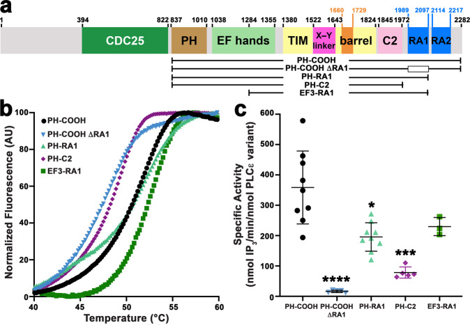
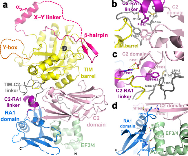
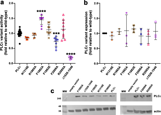
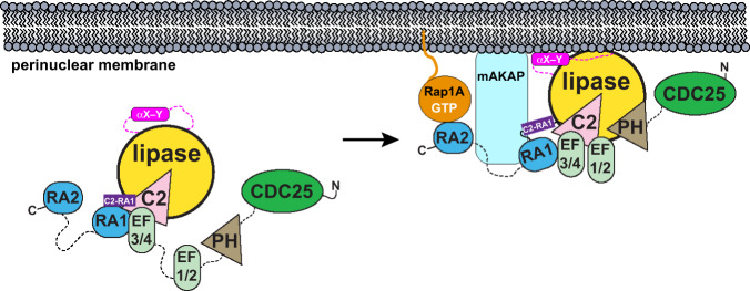
Phospholipase Cε (PLCε) generates lipid-derived second messengers at the plasma and perinuclear membranes in the cardiovascular system. It is activated in response to a wide variety of signals, such as those conveyed by Rap1A and Ras, through a mechanism that involves its C-terminal Ras association (RA) domains (RA1 and RA2). However, the complexity and size of PLCε has hindered its structural and functional analysis. Herein, we report the 2.7 Å crystal structure of the minimal fragment of PLCε that retains basal activity. This structure includes the RA1 domain, which forms extensive interactions with other core domains. A conserved amphipathic helix in the autoregulatory X-Y linker of PLCε is also revealed, which we show modulates activity in vitro and in cells. The studies provide the structural framework for the core of this critical cardiovascular enzyme that will allow for a better understanding of its regulation and roles in disease.


Conflict of interest statement

The authors declare no competing interests.

Figures

Fig. 1
Fig. 1. The PLCε RA domains have different roles in stability and basal activity.
a Domain structure of rat PLCε. The residue numbers above the diagram correspond to predicted (CDC25-EF1/2) or observed (6PMP, this study) domain boundaries. PLCε variants used in this study are diagrammed below. The open box in PLCε PH-COOH ΔRA1 corresponds to deletion of the RA1 domain. b Representative thermal denaturation curves of PH-COOH (black circles), PH-COOH ΔRA1 (blue inverted triangles), PH-RA1 (light green triangles), PH-C2 (purple diamonds), and EF3-RA1 (dark green squares). The most dramatic decreases in thermal stability are in variants that lack the RA1 domain. c Loss of both RA domains (PH-C2) or the RA1 domain (PH-COOH ΔRA1) decreases basal-specific activity up to ∼20-fold relative to PH-COOH, whereas deletion of RA2 (PH-RA1) only decreased activity ∼2-fold. Each data point shown represents the average of the duplicates from one technical repeat. Error bars reflect SD. Data for PLCε PH-COOH was previously reported. Significance was determined using a one-way ANOVA followed by Dunnett’s multiple comparisons test vs. PLCε PH-COOH. (****p  ≤ 0.0001, ***p ≤ 0.0005, **p ≤ 0.001, *p ≤ 0.05).
Fig. 2
Fig. 2. The structure of PLCε EF3-RA1 reveals an intimately associated C2-RA1 linker and RA1 domain.
a Crystal structure of PLCε EF3-RA1 with domains colored as in Fig. 1a. The catalytic Ca2+ is shown as a black sphere. The overall structure of the RA1 domain in the context of the crystal structure is similar to its solution structure (r.m.s.d. 1.4 Å for 73 Cα atoms, PDB ID 2BYE), with the greatest differences in the loop regions. Dashed lines correspond to disordered loops, and the N- and C-termini of the protein are labeled N and C, respectively. b F1982 in the C2-RA1 linker packs in a hydrophobic pocket formed by residues in the TIM barrel-C2 linker (gray) and the C2 domain. c R1987 further stabilizes interactions between the C2-RA1 linker and the C2 domain via a salt bridge with D1911. Dashed yellow lines correspond to hydrogen bonds or salt bridges ≤3.5 Å. d The RA1 domain interacts with both the C2 domain and the F3α helix of EF3/4. The C-terminus of the RA1 domain is labeled C.
Fig. 3
Fig. 3. Deletion of the αX–Y helix or mutation of the C2-RA1 linker alters lipase activity in cells.
COS-7 cells, which lack endogenous PLCε, were metabolically labeled with [3H]-myoinositol, transfected with PLCε variants, and the amount of [3H]-IPx quantified by scintillation counting. a Deletion of the αX–Y helix decreases lipase activity ∼5-fold relative to PLCε. The F2006A mutant, which helps stabilize the RA1-EF3/4 interface, had ∼1.4-fold lower activity. In contrast, the F1982A mutant, which disrupts the C2-RA1 linker, increased activity ∼1.5-fold. b Quantification of western blot data, showing that the differences in activity are not due to changes in expression. c Representative western blots, where empty vector and actin were used as the negative control and loading control, respectively. Each data point represents an individual experiment, and error bars reflect SD. Significance was determined using a one-way ANOVA followed by Dunnett’s multiple comparisons test vs. PLCε (****p ≤ 0.0001, ***p ≤ 0.0005, **p ≤ 0.001, *p ≤ 0.05).
Fig. 4
Fig. 4. Proposed model of basal PLCε regulation at the perinuclear membrane.
(Left) PLCε is present predominantly in the cytoplasm, and is maintained in a low-activity state by the autoinhibitory X–Y linker (hot pink) and the C2-RA1 linker (purple). The CDC25, PH, and RA2 domains, along with EF1/2, are not essential for full basal activity and may be flexibly connected to, or only transiently interact with, the PLCε core,,,. (Right) Localization of PLCε to the perinuclear membrane through interactions between the RA1 domain and the scaffolding protein mAKAP increase lipase activity,. RA1 binding to mAKAP could alter the conformation of, or displace, the C2-RA1 linker, increasing basal activity. Membrane association would also increase basal activity via interfacial activation, which may be facilitated by interactions between the αX–Y helix and the membrane or, alternatively, with other domains in PLCε or proteins at the target membrane, such as activated Rap1A.

References

    1. Kadamur G, Ross EM. Mammalian phospholipase C. Annu. Rev. Physiol. 2013;75:127–54. - PubMed
    1. Gresset A, Sondek J, Harden TK. The phospholipase C isozymes and their regulation. Subcell. Biochem. 2012;58:61–94. - PMC - PubMed
    1. Berridge MJ. The inositol trisphosphate/calcium signaling pathway in health and disease. Physiol. Rev. 2016;96:1261–96. - PubMed
    1. Fukami, K., Inanobe, S., Kanemaru, K. & Nakamura, Y. Phospholipase C is a key enzyme regulating intracellular calcium and modulating the phosphoinositide balance. Prog. Lipid Res.49, 429–437 (2010). - PubMed
    1. Oestreich EA, et al. Epac and phospholipase Cε regulate Ca2+ release in the heart by activation of protein kinase Cε and calcium-calmodulin kinase II. J. Biol. Chem. 2009;284:1514–22. - PMC - PubMed

Publication types

Substances

LinkOut - more resources