

Leishmania naiffi and lainsoni in French Guiana: Clinical features and phylogenetic variability
- PMID: 32797078
- PMCID: PMC7449503
- DOI: 10.1371/journal.pntd.0008380
Leishmania naiffi and lainsoni in French Guiana: Clinical features and phylogenetic variability
Abstract
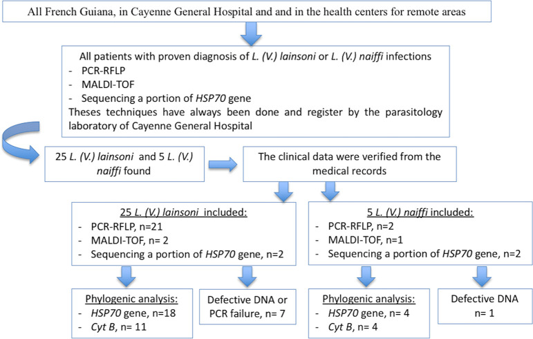
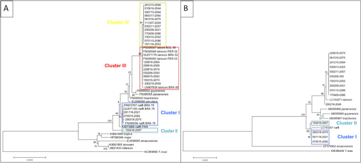
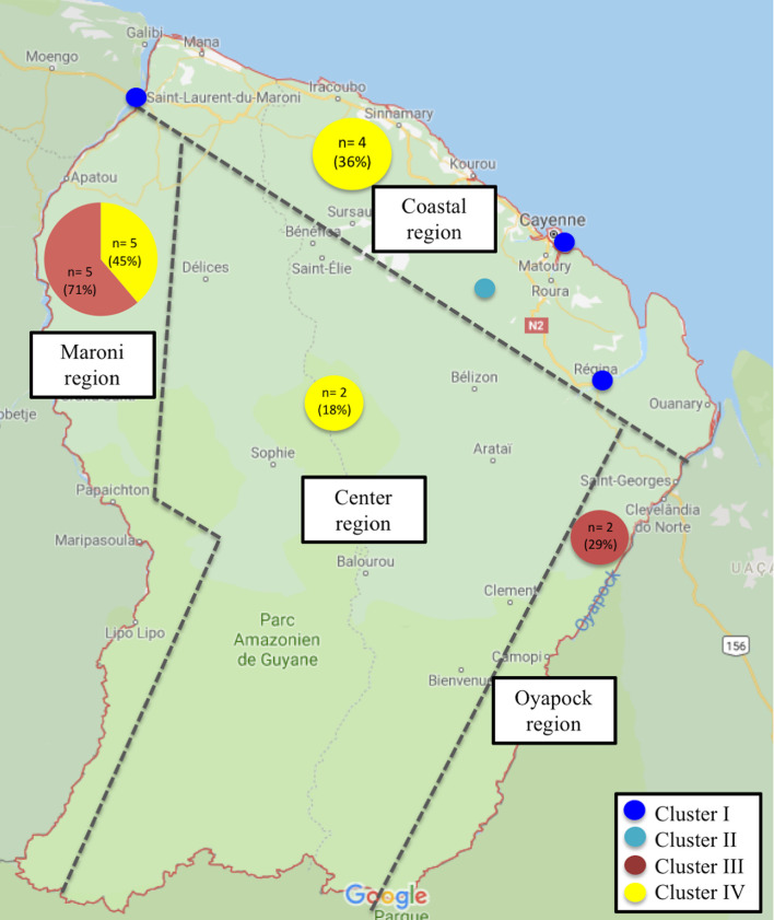
In French Guiana, five species are associated with Cutaneous Leishmaniasis (CL). Though infections with Leishmania guyanensis, L. (V.) braziliensis and L. (L.) amazonensis have been extensively described, there are few available clinical and genetic data on L. (V.) lainsoni and L. (V.) naiffi. We determined the clinical and epidemiological features of all cases of CL due to L. (V.) naiffi and L. (V.) lainsoni diagnosed in French Guiana between 2003 and 2019. Phylogenetic analysis was performed by sequencing a portion of HSP70 and cyt b genes. Five cases of L. naiffi and 25 cases of L. lainsoni were reported. Patients infected by L. (V.) lainsoni were usually infected on gold camps, mostly along the Maroni river (60%), while L. naiffi was observed in French patients infected on the coast (100%). A high number of pediatric cases (n = 5; 20%) was observed for L. (V.) lainsoni. A mild clinical course was observed for all cases of L. (V.) naiffi. HSP70 and cyt b partial nucleotide sequence analysis revealed different geographical clusters within L. (V.) naiffi and L. (V.) lainsoni but no association were found between phylogenetic and clinical features. Our data suggest distinct socio-epidemiological features for these two Leishmania species. Patients seem to get infected with L. (V.) naiffi during leisure activities in anthropized coastal areas, while L. (V.) lainsoni shares common features with L. (V.) guyanensis and braziliensis and seems to be acquired during professional activities in primary forest regions. Phylogenetic analysis has provided information on the intraspecific genetic variability of L. (V.) naiffi and L. (V.) lainsoni and how these genotypes are distributed at the geographic level.
Conflict of interest statement
The authors have declared that no competing interests exist.
Figures
References
-
- WHO | Leishmaniasis [Internet]. WHO. [cited 2019 sept 07]. Available from: http://www.who.int/leishmaniasis/en/
-
- Rotureau B, Couppié P, Nacher M, Dedet JP, Carme B. [Cutaneous leishmaniases in French Guiana]. Bull Soc Pathol Exot 1990. 2007. Oct;100(4):251–60. - PubMed
MeSH terms
Substances
LinkOut - more resources
Full Text Sources
