. 2020 Sep 18;48(16):9161-9180.
doi: 10.1093/nar/gkaa660.

FANCJ compensates for RAP80 deficiency and suppresses genomic instability induced by interstrand cross-links

Affiliations

FANCJ compensates for RAP80 deficiency and suppresses genomic instability induced by interstrand cross-links

Sanket Awate et al. Nucleic Acids Res. .

Abstract

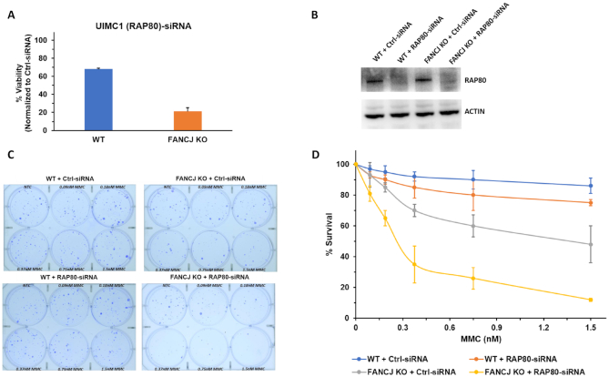
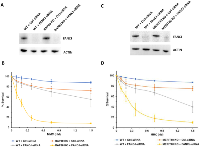
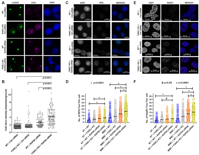
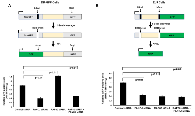
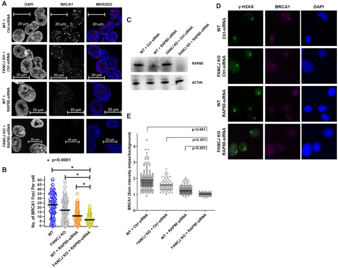
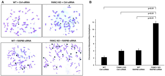
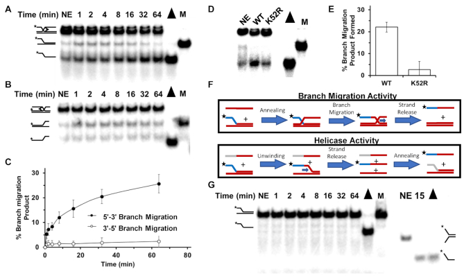
FANCJ, a DNA helicase and interacting partner of the tumor suppressor BRCA1, is crucial for the repair of DNA interstrand crosslinks (ICL), a highly toxic lesion that leads to chromosomal instability and perturbs normal transcription. In diploid cells, FANCJ is believed to operate in homologous recombination (HR) repair of DNA double-strand breaks (DSB); however, its precise role and molecular mechanism is poorly understood. Moreover, compensatory mechanisms of ICL resistance when FANCJ is deficient have not been explored. In this work, we conducted a siRNA screen to identify genes of the DNA damage response/DNA repair regime that when acutely depleted sensitize FANCJ CRISPR knockout cells to a low concentration of the DNA cross-linking agent mitomycin C (MMC). One of the top hits from the screen was RAP80, a protein that recruits repair machinery to broken DNA ends and regulates DNA end-processing. Concomitant loss of FANCJ and RAP80 not only accentuates DNA damage levels in human cells but also adversely affects the cell cycle checkpoint, resulting in profound chromosomal instability. Genetic complementation experiments demonstrated that both FANCJ's catalytic activity and interaction with BRCA1 are important for ICL resistance when RAP80 is deficient. The elevated RPA and RAD51 foci in cells co-deficient of FANCJ and RAP80 exposed to MMC are attributed to single-stranded DNA created by Mre11 and CtIP nucleases. Altogether, our cell-based findings together with biochemical studies suggest a critical function of FANCJ to suppress incompletely processed and toxic joint DNA molecules during repair of ICL-induced DNA damage.


Figures

Figure 1.
Figure 1.
Loss of RAP80 sensitizes FANCJ KO cells to a low dose of Mitomycin C. Panel (A), RNAi screening performed as described in Materials and Methods demonstrated that UIMC1 (RAP80) was one of the ten positive hits of the 240 DNA damage response genes that in RNAi-depleted FANCJ KO cells sensitized them to 6.25 nM MMC as measured by a cell viability assay. Panel (B), Western blot showing RNAi-mediated RAP80 depletion in the isogenic FANCJ KO and WT cell lines. Panel (C), WT or FANCJ KO cells were transfected with 30 nM control siRNA or RAP80 pool siRNA. After 24 h cells were re-transfected with 30 nM control siRNA or RAP80 pool siRNA, and 500–1000 cells were seeded in six-well plates with media containing the specified MMC concentration. Cells were incubated for 6 days after which media was changed. Colonies were detected 14 days after the initial seeding using crystal violet staining. Panel (D), Quantification of colony formation from three independent experiments.
Figure 2.
Figure 2.
RAP80 KO cells or Merit40 KO cells depleted of FANCJ are hypersensitive to MMC. Panels (A) and (C), western blot showing RNAi-mediated FANCJ depletion in the isogenic RAP80 KO or Merit40 KO and WT cell lines. Panels (B) and (D), WT and RAP80 KO or Merit40 KO cells were transfected with 30 nM control siRNA or FANCJ siRNA. After 24 h cells were re-transfected with 30 nM control siRNA or FANCJ siRNA, and 500–1000 cells were seeded in 6-well plates with media containing the specified MMC concentration. Cells were incubated for 6 days after which media was changed. Colonies were detected 14 days after the initial seeding using crystal violet staining. Quantification of colony formation is from three independent experiments.
Figure 3.
Figure 3.
Dual loss of FANCJ and RAP80 results in ssDNA accumulation at ICL-induced DNA damage, and elevated RPA and RAD51 foci in MMC-treated cells. Panels (A) and (B), Pso-ICL CldU ssDNA accumulation. WT or FANCJ KO cells were transfected with 30 nM control siRNA or RAP80 siRNA#1. After 24 h cells were re-transfected with 30 nM control siRNA or RAP80 siRNA#1. Cells were kept in 20 μM CldU for 48 hr. Forty-eight hr after the first transfection, cells were incubated with psoralen followed by laser treatment (1%) to induce Pso-ICLs. Cells were fixed and stained with anti-γ-H2AX and anti-CldU antibody. Panel (A), representative images of γ−H2AX and CldU stripes. Panel (B), The mean intensity of CldU localized to ICL stripes were quantified in single cells at 150 min after laser treatment. Panels (CF), Number of RPA and RAD51 foci per cell are elevated in the non-treated and MMC-treated cells doubly deficient in RAP80 and FANCJ. WT or FANCJ KO cells were transfected with 30 nM control siRNA or RAP80 siRNA#1. After 24 hr cells were re-transfected with 30 nM control siRNA or RAP80 siRNA#1. Forty-eight hours after the first transfection, cells were either left untreated or were treated with 3 μM MMC for 1 h and allowed to recover in fresh media. Sixteen hours later, RPA (Panels C, D) and Rad51 foci (Panels E, F) were determined after removing the soluble proteins and fixing chromatin bound proteins. Shown are representative images of the MMC-treated cells and quantitative analyses of both the untreated and MMC-treated cells. Over 150 cells were examined, and the results are mean ± standard error of mean (s.e.m.) from three biological repeats, with P-values determined by Student's t-tests.
Figure 4.
Figure 4.
Cells depleted of FANCJ and RAP80 display reduced double-strand break repair by homologous recombination or nonhomologous end-joining. The I-SceI DSB repair assay was used to measure HR repair or NHEJ of the indicated RNAi-depleted cell lines as described in Materials and Methods. Panel (A), HR efficiency in siRNA transfected DR-GFP cells. Panel (B), NHEJ efficiency in siRNA transfected EJ5 cells. The results are mean ± standard error of mean (s.e.m.) from three biological repeats, with P-values determined by Student's t-tests.
Figure 5.
Figure 5.
FANCJ and RAP80 act upstream and independently to elicit BRCA1 foci during the cellular response to mitomycin C and recruitment of BRCA1 to laser-induced psoralen cross-links. Panel (A), WT or FANCJ KO cells were transfected with 30 nM control siRNA or RAP80 siRNA#1. After 24 h, cells were re-transfected with 30 nM control siRNA or RAP80 siRNA#1. Forty-eight hr after the first transfection, cells were treated with 3 μM MMC for 1 h and allowed to recover in fresh media. Sixteen hours later, BRCA1 foci were examined after removing the soluble proteins and fixing chromatin bound proteins. Representative images for BRCA1 foci in the indicated cell lines are shown. Panel (B), Quantitative analysis of MMC-induced BRCA1 foci per cell. Over 100 cells were examined, and the results are mean ± standard error of mean (s.e.m.) from three biological repeats, with P-values determined by Student's t-tests. Panel (C), Western blot showing RNAi-mediated RAP80 depletion in the isogenic FANCJ KO or WT cell lines. Cells were incubated with psoralen followed by laser treatment (1%) to induce Pso-ICLs. Cells were fixed and stained with anti-γ−H2AX and anti-BRCA1 antibody Panel (D), representative images of γ−H2AX and BRCA1 stripes. Panel (E), Mean intensity of BRCA1 localized to ICL stripes were quantified in single cells at 20 min after laser treatment. The results are mean ± standard error of mean (s.e.m.) from three biological repeats, with P-values determined by Student's t-tests.
Figure 6.
Figure 6.
Elevated spontaneous chromosomal instability in cells deficient of FANCJ and RAP80. WT or FANCJ KO cells were treated with 30 nM control or RAP80 siRNA#1. After 24 h cells were re-transfected with 30 nM control or RAP80 siRNA#1. Forty-eight hr after the first transfection, cells were treated with 200 ng/ml of Colcemid for 3 hr and swollen in 75 mM KCl for 25 min at 37°C. Cells were fixed on ice with a 3:1 methanol/acetic acid solution. Metaphases were dropped onto slides preheated at 42°C, allowed to dry, and stained with Giemsa. The numbers of chromosomal abnormalities per metaphase were counted. Panel (A), Representative images of metaphase spreads. Panel (B), Quantification of chromosome abnormalities are mean ± standard error of mean (s.e.m.) from three independent experiments, with P-values determined by Student's t-tests.
Figure 7.
Figure 7.
FANCJ promotes three-stranded DNA branch-migration in the 5′ to 3′ direction. DNA products of branch-migration reaction mixtures containing a radiolabeled three-stranded DNA substrate FANCJ, and ATP as indicated in Materials and Methods were incubated for the indicated times and resolved by electrophoresis on non-denaturing 8% polyacrylamide gels and visualized by autoradiography. DNA substrates were designed to detect 5′ to 3′ branch-migration activity (Panel A) or 3′ to 5′ branch-migration activity (Panel B). Representative results from at least three independent experiments are shown. Quantitation of DNA products for Panels (A) and (B) is shown in Panel (C). Standard deviations are indicated by error bars. Panel (D), FANCJ-WT or FANCJ-K52R (ATPase-deficient mutant) was incubated with 5′ to 3′ branch-migratable substrate in presence of ATP for 30 min and products resolved by gel electrophoresis. Panel (E), Quantitation of at least three experiments from Panel (D). Panel (F), Depiction of experiments to distinguish whether helicase or branch-migration is responsible for the product observed in Panel (A). The ssDNA added to the branch-migratable substrate used in Panel (A) anneals to the upstream template strand (both entirely red). However, for the helicase assay the 5′ sequence (grey) of the ssDNA oligonucleotide was changed such that it can only anneal after the 2-stranded DNA substrate was unwound first. Panel (G), FANCJ-WT was incubated with the helicase substrate in the presence of ATP and a third partially complementary ssDNA oligonucleotide as depicted in the helicase activity schematic shown in Panel (F). Note duplex length of substrate on left is 63 bp; as a control for FANCJ catalytic activity, a 19 bp forked duplex substrate was tested for unwinding by FANCJ on the right.

References

    1. Hashimoto S., Anai H., Hanada K.. Mechanisms of interstrand DNA crosslink repair and human disorders. Genes Environ. 2016; 38:9. - PMC - PubMed
    1. Kohn K.W. Beyond DNA cross-linking: history and prospects of DNA-targeted cancer treatment–fifteenth Bruce F. Cain Memorial Award Lecture. Cancer Res. 1996; 56:5533–5546. - PubMed
    1. Deans A.J., West S.C.. DNA interstrand crosslink repair and cancer. Nat. Rev. Cancer. 2011; 11:467–480. - PMC - PubMed
    1. Pontel L.B., Rosado I.V., Burgos-Barragan G., Garaycoechea J.I., Yu R., Arends M.J., Chandrasekaran G., Broecker V., Wei W., Liu L. et al. .. Endogenous formaldehyde is a hematopoietic stem cell genotoxin and metabolic carcinogen. Mol. Cell. 2015; 60:177–188. - PMC - PubMed
    1. Voulgaridou G.P., Anestopoulos I., Franco R., Panayiotidis M.I., Pappa A.. DNA damage induced by endogenous aldehydes: current state of knowledge. Mutat. Res. 2011; 711:13–27. - PubMed

Publication types

MeSH terms