

The clinical utility of proton magnetic resonance spectroscopy in traumatic brain injury: recommendations from the ENIGMA MRS working group
- PMID: 32797399
- PMCID: PMC7882010
- DOI: 10.1007/s11682-020-00330-6
The clinical utility of proton magnetic resonance spectroscopy in traumatic brain injury: recommendations from the ENIGMA MRS working group
Abstract
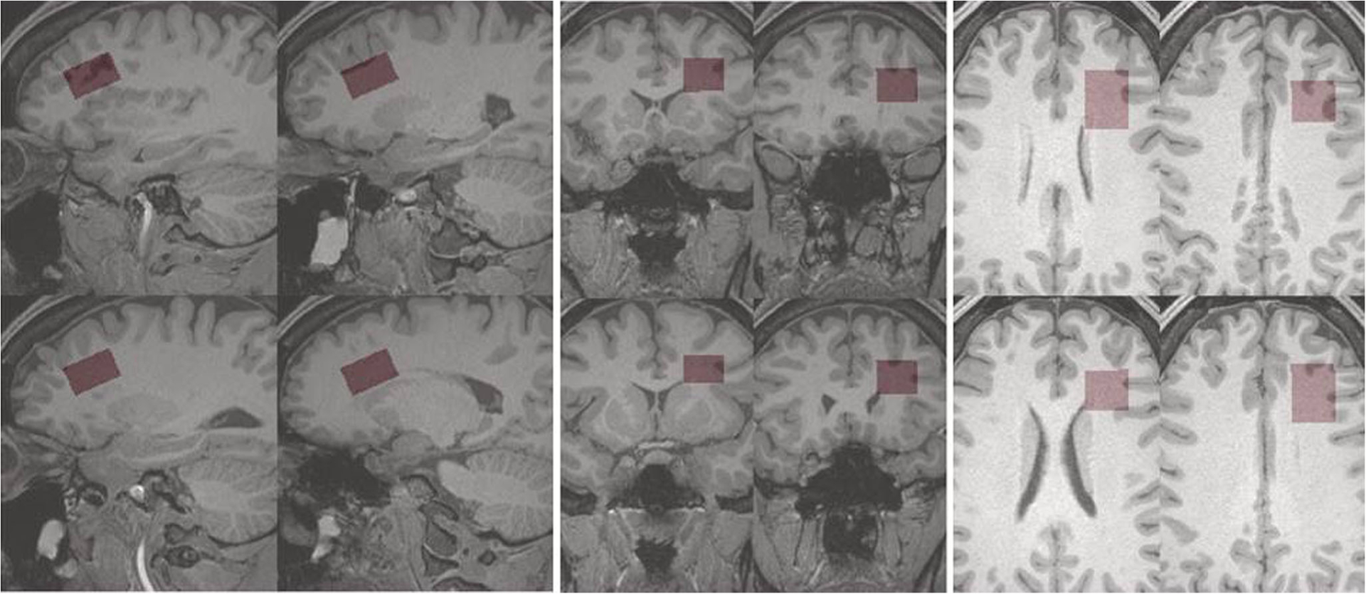
Proton (1H) magnetic resonance spectroscopy provides a non-invasive and quantitative measure of brain metabolites. Traumatic brain injury impacts cerebral metabolism and a number of research groups have successfully used this technique as a biomarker of injury and/or outcome in both pediatric and adult TBI populations. However, this technique is underutilized, with studies being performed primarily at centers with access to MR research support. In this paper we present a technical introduction to the acquisition and analysis of in vivo 1H magnetic resonance spectroscopy and review 1H magnetic resonance spectroscopy findings in different injury populations. In addition, we propose a basic 1H magnetic resonance spectroscopy data acquisition scheme (Supplemental Information) that can be added to any imaging protocol, regardless of clinical magnetic resonance platform. We outline a number of considerations for study design as a way of encouraging the use of 1H magnetic resonance spectroscopy in the study of traumatic brain injury, as well as recommendations to improve data harmonization across groups already using this technique.
Keywords: Brain injury; Concussion; Magnetic resonance spectroscopy; Trauma.
Conflict of interest statement
Figures

References
-
- Aaen GS, Holshouser BA, Sheridan C, Colbert C, McKenney M, Kido D, & Ashwal S (2010). Magnetic resonance spectroscopy predicts outcomes for children with nonaccidental trauma. Pediatrics, 125(2), 295–303. - PubMed
-
- Alessandri B, Doppenberg E, Zauner A, Woodward J, Choi S, & Bullock R (1999). Evidence for time-dependent glutamate-mediated glycolysis in head-injured patients: A microdialysis study. Acta Neurochirurgica. Supplement, 75, 25–28. - PubMed
-
- Alosco ML, Tripodis Y, Rowland B, Chua AS, Liao H, Martin B, Jarnagin J, Chaisson CE, Pasternak O, Karmacharya S, Koerte IK, Cantu RC, Kowall NW, McKee AC, Shenton ME, Greenwald R, McClean M, Stern RA, & Lin A (2019). A magnetic resonance spectroscopy investigation in symptomatic former NFL players. Brain Imaging and Behavior. 10.1007/s11682-019-00060-4. - DOI - PMC - PubMed
-
- Ariza M, Junqué C, Mataró M, Poca MA, Bargalló N, Olondo M, & Sahuquillo J (2004). Neuropsychological correlates of basal ganglia and medial temporal lobe NAA/Cho reductions in traumatic brain injury. Archives of Neurology, 61(4), 541–544. - PubMed
-
- Ashwal S, Holshouser BA, Shu SK, Simmons PL, Perkin RM, Tomasi LG, Knierim DS, Sheridan C, Craig K, Andrews GH, & Hinshaw DB (2000). Predictive value of proton magnetic resonance spectroscopy in pediatric closed head injury. Pediatric Neurology, 23(2), 114–125. - PubMed
MeSH terms
Grants and funding
- P41 EB015922/EB/NIBIB NIH HHS/United States
- R01 EB016064/EB/NIBIB NIH HHS/United States
- R21NS112853/NH/NIH HHS/United States
- R01 HD061504/HD/NICHD NIH HHS/United States
- U54EB020403/NH/NIH HHS/United States
- K99 NS096116/NS/NINDS NIH HHS/United States
- R21 NS112853/NS/NINDS NIH HHS/United States
- R01MH111671/NH/NIH HHS/United States
- R01MH116147/NH/NIH HHS/United States
- R01EB016064/NH/NIH HHS/United States
- R01 NS086885/NS/NINDS NIH HHS/United States
- P30AG008051/NH/NIH HHS/United States
- U01 NS093334/NS/NINDS NIH HHS/United States
- P30 AG008051/AG/NIA NIH HHS/United States
- R01 NS100952/NS/NINDS NIH HHS/United States
- R01 MH111671/MH/NIMH NIH HHS/United States
- P30 AG072980/AG/NIA NIH HHS/United States
- R01 MH116147/MH/NIMH NIH HHS/United States
- R56AG058854/NH/NIH HHS/United States
- K99NS096116/NH/NIH HHS/United States
- R01HD061504/NH/NIH HHS/United States
- U54 EB020403/EB/NIBIB NIH HHS/United States
- R01 NS097494/NS/NINDS NIH HHS/United States
- P41EB017183/NH/NIH HHS/United States
- R56 AG058854/AG/NIA NIH HHS/United States
- R01NS097494/National Institutes of Health (US)
- P41EB015922/NH/NIH HHS/United States
- P41 EB017183/EB/NIBIB NIH HHS/United States
LinkOut - more resources
Full Text Sources
Medical
Miscellaneous
