

A Case of Spontaneous Pneumothorax 21 Days After Diagnosis of Coronavirus Disease 2019 (COVID-19) Pneumonia
- PMID: 32798215
- PMCID: PMC7447295
- DOI: 10.12659/AJCR.925787
A Case of Spontaneous Pneumothorax 21 Days After Diagnosis of Coronavirus Disease 2019 (COVID-19) Pneumonia
Abstract
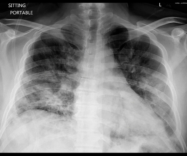
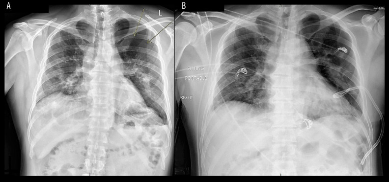
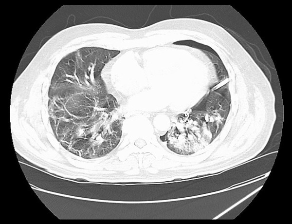
BACKGROUND At the end of 2019, coronavirus (SARS-CoV-2) was recognized as the cause of a cluster of pneumonia cases in Wuhan, a city in China. There are numerous complications associated with COVID-19 infection, such as acute respiratory distress syndrome, renal failure, circulatory shock, and multi-organ failure. Spontaneous pneumothorax following COVID-19 pneumonia is an extremely rare complication. CASE REPORT We report the case of a 49-year-old man with a past medical history of type 2 diabetes mellitus with an initial presentation of cough, shortness of breath, and fever. He was diagnosed with COVID-19 pneumonia and rapidly deteriorated on the day of admission, requiring initiation of mechanical ventilation. The patient recovered clinically and was discharged home. He returned 21 days after discharge with a spontaneous pneumothorax. CONCLUSIONS Spontaneous pneumothorax is a rare complication after apparent recovery from COVID-19 pneumonia. It is imperative that treating physicians are aware of this complication in order to recognize it early and treat it promptly.
Conflict of interest statement
None.
Figures
Similar articles
-
The coronavirus diseases 2019 (COVID-19) pneumonia with spontaneous pneumothorax: a case report.BMC Infect Dis. 2020 Sep 9;20(1):662. doi: 10.1186/s12879-020-05384-x. BMC Infect Dis. 2020. PMID: 32907540 Free PMC article.
-
Pneumomediastinum and spontaneous pneumothorax as an extrapulmonary complication of COVID-19 disease.Emerg Radiol. 2020 Dec;27(6):727-730. doi: 10.1007/s10140-020-01806-0. Epub 2020 Jun 11. Emerg Radiol. 2020. PMID: 32524296 Free PMC article.
-
Pneumothorax in a COVID-19 Pneumonia Patient without Underlying Risk Factors.Intern Med. 2020 Nov 15;59(22):2921-2925. doi: 10.2169/internalmedicine.5731-20. Epub 2020 Oct 7. Intern Med. 2020. PMID: 33028774 Free PMC article.
-
Spontaneous pneumomediastinum, pneumothorax and subcutaneous emphysema in COVID-19: case report and literature review.Rev Inst Med Trop Sao Paulo. 2020 Oct 9;62:e76. doi: 10.1590/S1678-9946202062076. eCollection 2020. Rev Inst Med Trop Sao Paulo. 2020. PMID: 33053145 Free PMC article. Review.
-
Case Report: COVID-19-Related Pneumothorax-Case Series Highlighting a Significant Complication.Am J Trop Med Hyg. 2020 Sep;103(3):1166-1169. doi: 10.4269/ajtmh.20-0713. Am J Trop Med Hyg. 2020. PMID: 32662394 Free PMC article. Review.
Cited by
-
Pneumothorax due to COVID-19: Analysis of case reports.Respir Med Case Rep. 2021;34:101490. doi: 10.1016/j.rmcr.2021.101490. Epub 2021 Jul 26. Respir Med Case Rep. 2021. PMID: 34336592 Free PMC article.
-
Applying Parse's Human Becoming Theory for Caring of an Elderly with Spontaneous Pneumothorax Following the COVID-19: A Case Study.J Caring Sci. 2023 Oct 10;13(1):3-11. doi: 10.34172/jcs.2023.33017. eCollection 2024 Feb. J Caring Sci. 2023. PMID: 38659434 Free PMC article.
-
Respiratory Complications after COVID-19.Oman Med J. 2022 Jan 31;37(1):e343. doi: 10.5001/omj.2022.52. eCollection 2022 Jan. Oman Med J. 2022. PMID: 35282425 Free PMC article. Review.
-
Clinical outcomes of pleural drainage on pneumothorax and hydrothorax in critically ill patients with COVID-19: A case series with literature review.Heart Lung. 2021 Mar-Apr;50(2):213-219. doi: 10.1016/j.hrtlng.2020.12.007. Epub 2020 Dec 9. Heart Lung. 2021. PMID: 33310504 Free PMC article. Review.
-
Cardio-Pulmonary Sequelae in Recovered COVID-19 Patients: Considerations for Primary Care.J Prim Care Community Health. 2021 Jan-Dec;12:21501327211023726. doi: 10.1177/21501327211023726. J Prim Care Community Health. 2021. PMID: 34096390 Free PMC article. Review.
References
-
- World Health Organization (WHO) https://www.who.int/emergencies/diseases/novel-coronavirus-2019.
Publication types
MeSH terms
LinkOut - more resources
Full Text Sources
Medical
Miscellaneous
