. 2020 Oct 1;222(9):1462-1467.
doi: 10.1093/infdis/jiaa507.

Methods of Inactivation of SARS-CoV-2 for Downstream Biological Assays

Affiliations

Methods of Inactivation of SARS-CoV-2 for Downstream Biological Assays

Edward I Patterson et al. J Infect Dis. .

Abstract

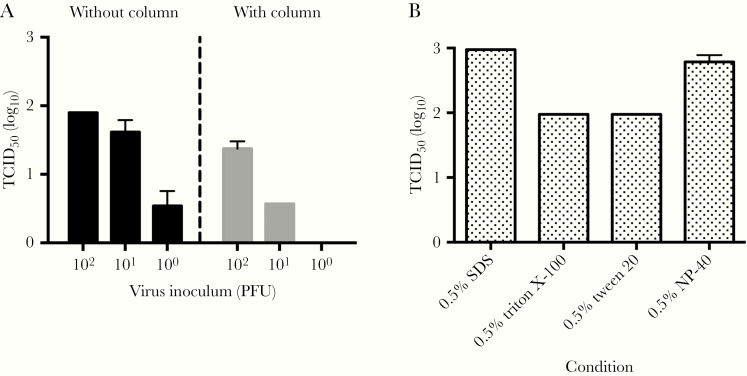
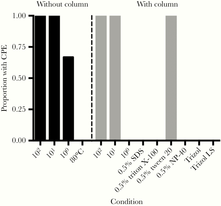
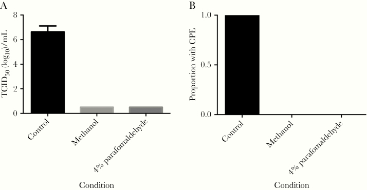
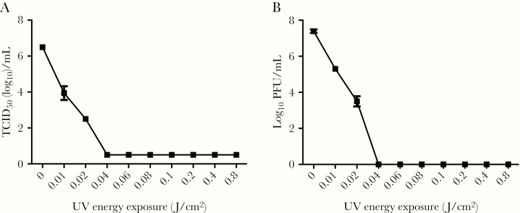
The scientific community has responded to the coronavirus disease 2019 (COVID-19) pandemic by rapidly undertaking research to find effective strategies to reduce the burden of this disease. Encouragingly, researchers from a diverse array of fields are collectively working towards this goal. Research with infectious severe acute respiratory syndrome coronavirus 2 (SARS-CoV-2) is undertaken in high-containment laboratories; however, it is often desirable to work with samples at lower-containment levels. To facilitate the transfer of infectious samples from high-containment laboratories, we have tested methods commonly used to inactivate virus and prepare the sample for additional experiments. Incubation at 80°C, a range of detergents, Trizol reagents, and UV energies were successful at inactivating a high titer of SARS-CoV-2. Methanol and paraformaldehyde incubation of infected cells also inactivated the virus. These protocols can provide a framework for in-house inactivation of SARS-CoV-2 in other laboratories, ensuring the safe use of samples in lower-containment levels.

Keywords: SARS-Cov-2; Trizol; detergents; inactivation; methanol; paraformaldehyde; temperature.


Figures

Figure 1.
Figure 1.
Limit of detection of SARS-CoV-2 by different methods. A, Assay to quantify the limits of detection. Known titers of virus were prepared with (right) or without (left) concentrating the sample in a centrifugal column. Quantification of controls was performed using TCID50. Lower limit of detection was 3.16 TCID50/mL: (n = 3; mean ± SEM). B, Cytotoxicity from uninfected control samples diluted in a TCID50 assay (n = 3; mean ± SEM). Abbreviations: PFU, plaque-forming unit; SARS-CoV-2, severe acute respiratory syndrome coronavirus 2; SDS, sodium dodecyl sulfate; SEM, standard error of the mean; TCID50, 50% tissue culture infectious dose.
Figure 2.
Figure 2.
The proportion of SARS-CoV-2 inactivation assays with CPE. Samples were either diluted for assays (left) or inactivation agent removed using centrifugal columns (right). Control samples with 100, 101, and 102 PFU of SARS-CoV-2 were used as positive controls and to determine the limit of detection for each method. Abbreviations: CPE, cytopathic effect; PFU, plaque-forming unit; SARS-CoV-2, severe acute respiratory syndrome coronavirus 2; SDS, sodium dodecyl sulfate.
Figure 3.
Figure 3.
Effect of fixation on infectious virus in cells. A, TCID50 assay comparing untreated infected cells with fixed infected cells. Lower limit of detection was 0.5 TCID50 (log10)/mL (n = 3; mean ± SEM). B, The proportion of inactivation assays with CPE. Abbreviations: CPE, cytopathic effect; SEM, standard error of the mean; TCID50, 50% tissue culture infectious dose.
Figure 4.
Figure 4.
Quantification of SARS-CoV-2 following exposure to different energies of UV light. A, The concentration of viable SARS-CoV-2 following exposure to UV light measured by TCID50 assay. Lower limit of detection was 0.5 TCID50(log10)/mL. B, The concentration of viable SARS-CoV-2 following exposure to UV measured by plaque assay. Both assays confirmed the complete inactivation of the sample after exposure to 0.04 J/cm2 or higher UV energy. Results are expressed as mean ± SEM (n = 3). Abbreviations: PFU, plaque-forming unit; SEM, standard error of the mean; TCID50, 50% tissue culture infectious dose.

Update of

References

    1. Wu F, Zhao S, Yu B, et al. A new coronavirus associated with human respiratory disease in China. Nature 2020; 579:265–9. - PMC - PubMed
    1. Zhou P, Yang XL, Wang XG, et al. A pneumonia outbreak associated with a new coronavirus of probable bat origin. Nature 2020; 579:270–3. - PMC - PubMed
    1. World Health Organization. WHO Coronavirus disease (COVID-19) dashboard https://covid19.who.int. Accessed 13 August 2020.
    1. Lu R, Zhao X, Li J, et al. Genomic characterisation and epidemiology of 2019 novel coronavirus: implications for virus origins and receptor binding. Lancet 2020; 395:565–74. - PMC - PubMed
    1. Patterson EI, Warmbrod KL, Bouyer DH, Forrester NL. Evaluation of the inactivation of Venezuelan equine encephalitis virus by several common methods. J Virol Methods 2018; 254:31–4. - PMC - PubMed

Publication types