

Protein expression of angiotensin-converting enzyme 2, a SARS-CoV-2-specific receptor, in fetal and placental tissues throughout gestation: new insight for perinatal counseling
- PMID: 32798244
- PMCID: PMC7461228
- DOI: 10.1002/uog.22178
Protein expression of angiotensin-converting enzyme 2, a SARS-CoV-2-specific receptor, in fetal and placental tissues throughout gestation: new insight for perinatal counseling
Abstract
Objective: Pregnant women can be infected by severe acute respiratory syndrome coronavirus 2 (SARS-CoV-2), yet the incidence of perinatal infection is low. We hypothesized that this could be related to low expression of the membrane receptor for SARS-CoV-2, angiotensin-converting enzyme 2 (ACE2), in the fetoplacental unit. We evaluated protein expression of ACE2 at various gestational ages in both placentae and fetal organs from pregnancies not infected with SARS-CoV-2.
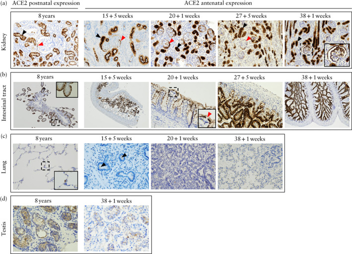
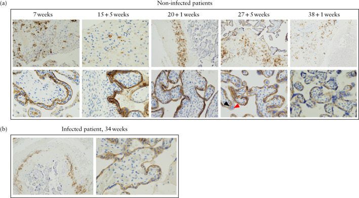
Methods: In May 2020, using samples from a registered biobank, we performed immunohistochemical analysis for ACE2 in tissue samples from fetal organs and placentae from five cases of second- or third-trimester medical termination of pregnancy in healthy women (performed between 15 and 38 weeks' gestation), as well as a further two placentae, one from a 7-week spontaneous miscarriage in a non-infected woman and one from a symptomatic pregnant woman positive for SARS-CoV-2 delivered by Cesarean section at 34 weeks. Samples were paraffin-embedded and organ tissues included kidney, brain, lung, intestinal tract, heart and testis. Matching tissues (kidney, intestinal tract, lung and testis) from autopsies of four 8-year-old children were tested as controls. Tissue sections were incubated with rabbit monoclonal anti-ACE2, and protein expression of ACE2 was detected by immunohistochemistry.
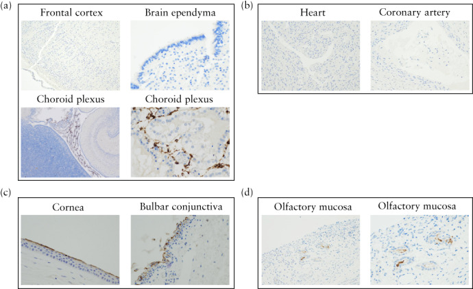
Results: ACE2 expression was detected in fetal kidney, rectum and ileum samples from 15 weeks onwards and in the pediatric controls. It was barely detectable in fetal lung samples at 15 + 5 weeks' gestation and not detectable thereafter, and, in the pediatric controls, ACE2 was detectable only in type-2 pneumocytes. No ACE2 expression was found in the cerebral ependymal or parenchymal tissues or in cardiac tissues. ACE2 was expressed in placental syncytiotrophoblast and cytotrophoblast samples, but not in the amnion, from 7 weeks onwards. The intensity and distribution of ACE2 staining in the placenta from the symptomatic SARS-CoV-2 woman was similar to that in the non-infected placentae.
Conclusions: Marked placental expression of ACE2 provides a rationale for vertical transmission at the cellular level. Absence of ACE2 expression in the fetal brain and heart is reassuring regarding the risk of congenital malformation. Clinical follow-up of infected pregnant women and their children is needed to validate these observations. © 2020 International Society of Ultrasound in Obstetrics and Gynecology.
Keywords: ACE2; COVID-19; SARS-CoV-2; fetal organs; placenta; protein expression; vertical transmission.
© 2020 International Society of Ultrasound in Obstetrics and Gynecology.
Figures
Comment in
-
Obstetric and neonatal literature is complex and should be merged to understand perinatal SARS-CoV-2 infection.Ultrasound Obstet Gynecol. 2021 Feb;57(2):351-352. doi: 10.1002/uog.23582. Ultrasound Obstet Gynecol. 2021. PMID: 33524234 Free PMC article.
References
-
- Kimberlin DW, Stagno S. Can SARS‐CoV‐2 Infection Be Acquired In Utero?: More Definitive Evidence Is Needed. JAMA 2020; 323: 1788–1789. - PubMed
MeSH terms
Substances
LinkOut - more resources
Full Text Sources
Other Literature Sources
Miscellaneous
