

Exposure to prenatal PCBs shifts the timing of neurogenesis in the hypothalamus of developing rats
- PMID: 32798281
- PMCID: PMC7812377
- DOI: 10.1002/jez.2404
Exposure to prenatal PCBs shifts the timing of neurogenesis in the hypothalamus of developing rats
Abstract
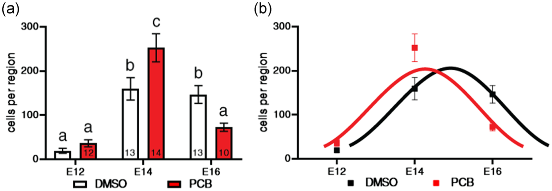
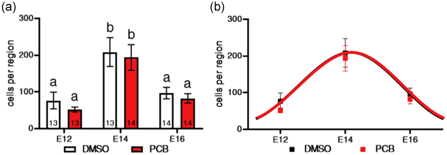
The developing brain is highly sensitive to the hormonal milieu, with gonadal steroid hormones involved in neurogenesis, neural survival, and brain organization. Limited available evidence suggests that endocrine-disrupting chemicals (EDCs) may perturb these developmental processes. In this study, we tested the hypothesis that prenatal exposure to a mixture of polychlorinated biphenyls (PCBs), Aroclor 1221, would disrupt the normal timing of neurogenesis in two hypothalamic regions: the ventromedial nucleus (VMN) and the preoptic area (POA). These regions were selected because of their important roles in the control of sociosexual behaviors that are perturbed in adulthood by prenatal EDC exposure. Pregnant Sprague-Dawley rats were exposed to PCBs from Embryonic Day 8 (E8) to E18, encompassing the period of neurogenesis of all hypothalamic neurons. To determine the birth dates of neurons, bromo-2-deoxy-5-uridine (BrdU) was administered to dams on E12, E14, or E16. On the day after birth, male and female pups were perfused, brains immunolabeled for BrdU, and numbers of cells counted. In the VMN, exposure to PCBs significantly advanced the timing of neurogenesis compared to vehicle-treated pups, without changing the total number of BrdU+ cells. In the POA, PCBs did not change the timing of neurogenesis nor the total number of cells born. This is the first study to show that PCBs can shift the timing of neurogenesis in the hypothalamus, specifically in the VMN but not the POA. This result has implications for functions controlled by the VMN, especially sociosexual behaviors, as well as for sexual selection more generally.
Keywords: Aroclor 1221; endocrine-disrupting chemical; hypothalamus; neurogenesis; polychlorinated biphenyl; preoptic area; ventromedial nucleus.
© 2020 Wiley Periodicals LLC.
Figures


Similar articles
-
Endocrine-disrupting chemicals alter the neuromolecular phenotype in F2 generation adult male rats.Physiol Behav. 2019 Nov 1;211:112674. doi: 10.1016/j.physbeh.2019.112674. Epub 2019 Sep 3. Physiol Behav. 2019. PMID: 31491443 Free PMC article.
-
Social and neuromolecular phenotypes are programmed by prenatal exposures to endocrine-disrupting chemicals.Mol Cell Endocrinol. 2019 Jan 5;479:133-146. doi: 10.1016/j.mce.2018.09.010. Epub 2018 Oct 1. Mol Cell Endocrinol. 2019. PMID: 30287398 Free PMC article.
-
Prenatal PCBs disrupt early neuroendocrine development of the rat hypothalamus.Toxicol Appl Pharmacol. 2011 Apr 1;252(1):36-46. doi: 10.1016/j.taap.2011.01.012. Epub 2011 Jan 26. Toxicol Appl Pharmacol. 2011. PMID: 21277884 Free PMC article.
-
Endocrine-disrupting chemicals: Effects on neuroendocrine systems and the neurobiology of social behavior.Horm Behav. 2019 May;111:7-22. doi: 10.1016/j.yhbeh.2018.11.006. Epub 2018 Dec 4. Horm Behav. 2019. PMID: 30476496 Free PMC article. Review.
-
Early developmental actions of endocrine disruptors on the hypothalamus, hippocampus, and cerebral cortex.J Toxicol Environ Health B Crit Rev. 2011;14(5-7):328-45. doi: 10.1080/10937404.2011.578556. J Toxicol Environ Health B Crit Rev. 2011. PMID: 21790315 Free PMC article. Review.
Cited by
-
Neuroendocrine and Developmental Impacts of Early Life Exposure to EDCs.J Endocr Soc. 2024 Dec 10;9(1):bvae195. doi: 10.1210/jendso/bvae195. eCollection 2024 Nov 26. J Endocr Soc. 2024. PMID: 39659541 Free PMC article.
-
Perinatal phthalate exposure increases developmental apoptosis in the rat medial prefrontal cortex.Neurotoxicology. 2021 Dec;87:167-173. doi: 10.1016/j.neuro.2021.09.007. Epub 2021 Sep 29. Neurotoxicology. 2021. PMID: 34599995 Free PMC article.
-
Exposure to environmental chemicals and perinatal psychopathology.Biochem Pharmacol. 2022 Jan;195:114835. doi: 10.1016/j.bcp.2021.114835. Epub 2021 Nov 11. Biochem Pharmacol. 2022. PMID: 34774531 Free PMC article. Review.
-
Transcriptomic analysis of effects of developmental PCB exposure in the hypothalamus of female rats.Mol Cell Endocrinol. 2025 Apr 1;599:112460. doi: 10.1016/j.mce.2025.112460. Epub 2025 Jan 9. Mol Cell Endocrinol. 2025. PMID: 39798907 Free PMC article.
-
Effects of endocrine-disrupting chemicals on hypothalamic oxytocin and vasopressin systems.J Exp Zool A Ecol Integr Physiol. 2022 Jan;337(1):75-87. doi: 10.1002/jez.2475. Epub 2021 May 21. J Exp Zool A Ecol Integr Physiol. 2022. PMID: 34018699 Free PMC article.
References
-
- Bellanger M, Demeneix B, Grandjean P, Zoeller RT, & Trasande L (2015). Neurobehavioral deficits, diseases, and associated costs of exposure to endocrine-disrupting chemicals in the European Union. Journal of Clinical Endocrinology and Metabolism, 100(4), 1256–1266. 10.1210/jc.2014-4323 - DOI - PMC - PubMed
Publication types
MeSH terms
Substances
Grants and funding
LinkOut - more resources
Full Text Sources
