. 2020 Sep 9;28(3):465-474.e4.
doi: 10.1016/j.chom.2020.07.018. Epub 2020 Jul 30.

Replication-Competent Vesicular Stomatitis Virus Vaccine Vector Protects against SARS-CoV-2-Mediated Pathogenesis in Mice

Affiliations

Replication-Competent Vesicular Stomatitis Virus Vaccine Vector Protects against SARS-CoV-2-Mediated Pathogenesis in Mice

James Brett Case et al. Cell Host Microbe. .

Abstract

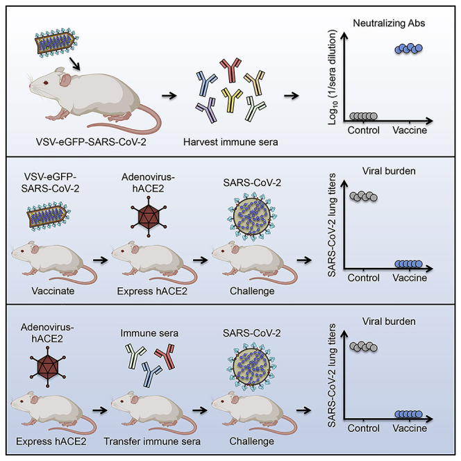
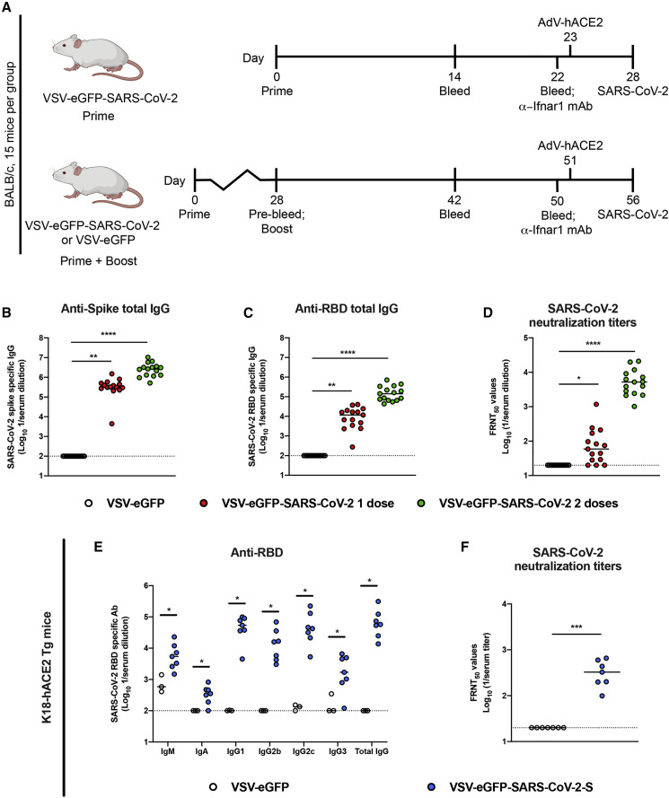
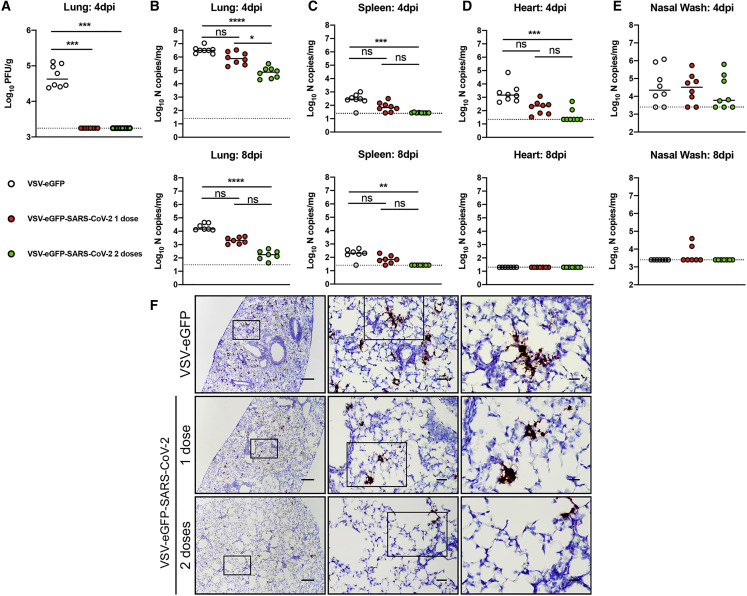
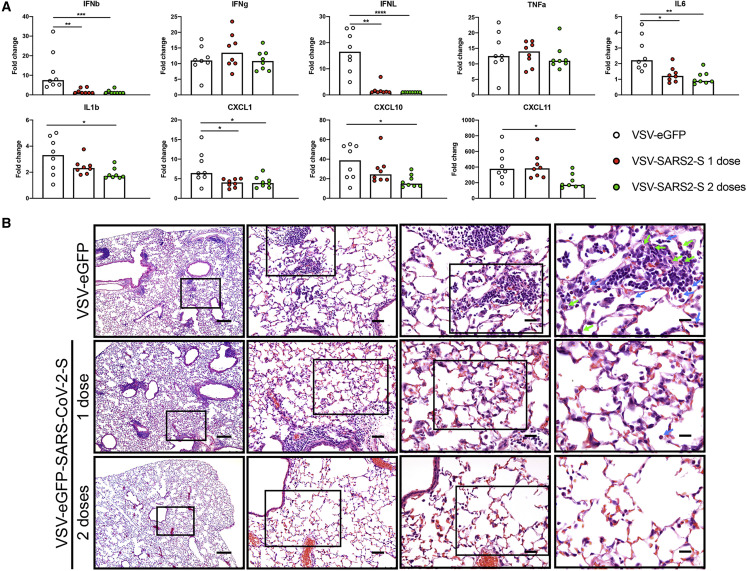
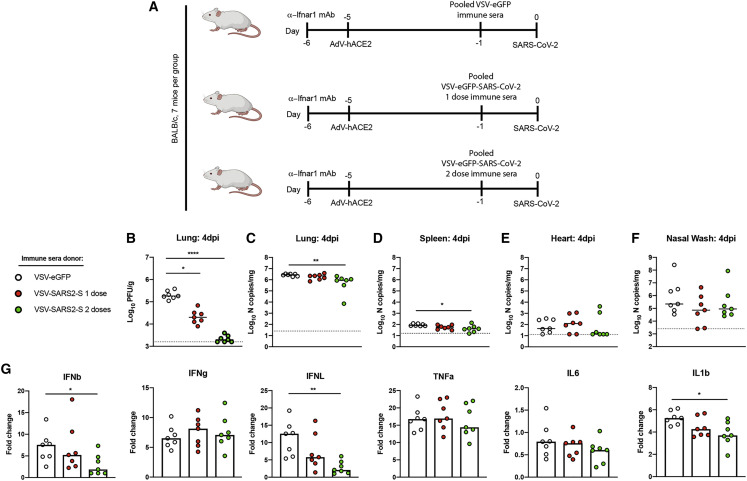
Severe acute respiratory syndrome coronavirus 2 (SARS-CoV-2) has caused millions of human infections, and an effective vaccine is critical to mitigate coronavirus-induced disease 2019 (COVID-19). Previously, we developed a replication-competent vesicular stomatitis virus (VSV) expressing a modified form of the SARS-CoV-2 spike gene in place of the native glycoprotein gene (VSV-eGFP-SARS-CoV-2). Here, we show that vaccination with VSV-eGFP-SARS-CoV-2 generates neutralizing immune responses and protects mice from SARS-CoV-2. Immunization of mice with VSV-eGFP-SARS-CoV-2 elicits high antibody titers that neutralize SARS-CoV-2 and target the receptor binding domain that engages human angiotensin-converting enzyme-2 (ACE2). Upon challenge with a human isolate of SARS-CoV-2, mice that expressed human ACE2 and were immunized with VSV-eGFP-SARS-CoV-2 show profoundly reduced viral infection and inflammation in the lung, indicating protection against pneumonia. Passive transfer of sera from VSV-eGFP-SARS-CoV-2-immunized animals also protects naive mice from SARS-CoV-2 challenge. These data support development of VSV-SARS-CoV-2 as an attenuated, replication-competent vaccine against SARS-CoV-2.

Keywords: COVID-19; SARS-CoV-2; correlates; humoral immunity; immunity; neutralizing antibodies; vaccine; vesicular stomatitis virus.


Conflict of interest statement

Declaration of Interests M.S.D. is a consultant for Inbios, Vir Biotechnology, and NGM Biopharmaceuticals and is on the Scientific Advisory Board of Moderna. M.J.H. is a member of the Data and Safety Monitoring Board for AstroZeneca and founder of NuPeak Therapeutics. The Diamond laboratory has received funding under sponsored research agreements from Moderna, Vir Biotechnology, and Emergent BioSolutions. The Whelan laboratory has received funding under sponsored research agreements from Vir Biotechnology. S.P.J.W., P.W.R., M.S.D., and J.B.C. have filed a disclosure with Washington University for the recombinant VSV.

Figures

None
Graphical abstract
Figure 1
Figure 1
Immunogenicity of VSV-eGFP-SARS-CoV-2 (A) Scheme of vaccination and SARS-CoV-2 challenge. (B–D) Four-week-old female BALB/c mice were immunized with VSV-eGFP or VSV-eGFP-SARS-CoV-2. Some of the immunized mice were boosted with their respective vaccines four weeks after primary vaccination. IgG responses in the sera of vaccinated mice were evaluated three weeks after priming or boosting by ELISA for binding to SARS-CoV-2 S (B) or RBD (C) or two weeks after priming or boosting by focus reduction neutralization test (FRNT) (D) (n = 15 per group; one-way ANOVA with Dunnett’s post-test: ∗∗∗∗p < 0.0001). Bars indicate median values. (E and F) Four-week-old K18-hACE2 transgenic mice were immunized with VSV-eGFP or VSV-eGFP-SARS-CoV-2 via an intranasal route. Three weeks later, serum was harvested and levels of anti-SARS-CoV-2 RBD antibodies (IgM, IgA, IgG1, IgG2b, IgG2c, IgG3, and total IgG) were determined by ELISA (n = 3–7 per group; Mann-Whitney test: p < 0.05) (E), or neutralizing antibody titers were determined by FRNT (F) (n = 7 per group; Mann-Whitney test: ∗∗∗p < 0.001). See Figure S1.
Figure 2
Figure 2
VSV-eGFP-SARS-CoV-2 Protects Mice against SARS-CoV-2 Infection (A–E) Three weeks after priming or boosting with VSV-eGFP or VSV-eGFP-SARS-CoV-2, immunized animals were treated with anti-Ifnar1 mAb and one day later, animals were transduced with 2.5 × 108 PFU of AdV-hACE2 by intranasal administration. Five days later, animals were challenged with 3 × 105 PFU of SARS-CoV-2 via intranasal administration. At 4 or 8 dpi tissues were harvested, and viral burden was determined in the lung ([A] and [B]), spleen (C), heart (D), and nasal washes (E) by plaque (A) or RT-qPCR ([B]–[E]) assay (n = 7–8 mice per group; Kruskal-Wallis test with Dunn’s post-test ([A]–[E]): ns, not significant, p < 0.05, ∗∗p < 0.01, ∗∗∗p < 0.001, ∗∗∗∗p < 0.0001). Dotted lines indicate the limit of detection. Bars indicate median values. (F) SARS-CoV-2 RNA in situ hybridization of lungs of mice vaccinated with VSV-eGFP or VSV-eGFP-SARS-CoV-2 and challenged with SARS-CoV-2 at 4 dpi. Images show 40×- (left; scale bars, 100 μm), 200×- (middle; scale bars, 100 μm), and 400×-magnification (right; scale bars, 10 μm; representative images from n = 3 lungs per group; 10 fields per slide).
Figure 3
Figure 3
VSV-eGFP-SARS-CoV-2 Protects Mice from SARS-CoV-2 Lung Inflammation (A) Lungs of VSV-eGFP- or VSV-eGFP-SARS-CoV-2-immunized mice were evaluated at 4 dpi for cytokine and chemokine expression by RT-qPCR assay. Data are shown as fold change in gene expression compared to fully naive, age-matched animals after normalization to Gapdh (n = 7–8 per group, Kruskal-Wallis test with Dunn’s post-test: p < 0.05, ∗∗p < 0.01, ∗∗∗p < 0.001, ∗∗∗∗p < 0.0001). Bars indicate median values. (B) Hematoxylin and eosin staining of lung sections from immunized mice at 8 dpi with SARS-CoV-2 (3 × 105 PFU). Images show 40×- (left; scale bars, 250 μm), 200×- (second from left; scale bars, 50 μm), 400×- (third from left; scale bars, 25 μm), and 630×-magnification (right; scale bars, 10 μm). Arrows indicate neutrophils in the alveolar septum (green) and airspace (blue). Representative images are shown from n = 3 lungs per group; 10 fields per slide).
Figure 4
Figure 4
Vaccine-Induced Sera Limits SARS-CoV-2 Infection (A) Passive transfer of immune sera and SARS-CoV-2 challenge scheme. Ten-week-old female BALB/c mice were treated with anti-Ifnar1 mAb, and one day later animals were transduced with 2.5 × 108 PFU of AdV-hACE2 by intranasal administration. Four days later, animals were administered 100 μL of pooled immune sera collected from VSV-eGFP or VSV-eGFP-SARS-CoV-2 vaccinated mice after one or two immunizations. One day later, animals were challenged with 3 × 105 PFU of SARS-CoV-2 via intranasal administration. (B–F) At 4 dpi tissues were harvested, and viral burden was determined in the lung ([B] and [C]), spleen (D), heart (E), and nasal washes (F) by plaque (B) or RT-qPCR ([C]–[F]) assays (n = 7 mice per group; Kruskal-Wallis test with Dunn’s post-test ([B]–[F]): p < 0.05, ∗∗p < 0.01, ∗∗∗∗p < 0.0001). Dotted lines indicate the limit of detection. (G) Lungs of mice treated with immune sera were evaluated at 4 dpi for cytokine expression by RT-qPCR assay. Data are shown as fold change in gene expression compared to naive, age-matched animals after normalization to Gapdh (n = 7 per group, Kruskal-Wallis test with Dunn’s post-test: p < 0.05, ∗∗p < 0.01). (BG) Bars indicate median values.

Update of

Comment in

  • Snatching the Crown from SARS-CoV-2.
    Coughlan L. Coughlan L. Cell Host Microbe. 2020 Sep 9;28(3):360-363. doi: 10.1016/j.chom.2020.08.007. Cell Host Microbe. 2020. PMID: 32910919 Free PMC article.

References

    1. Alsoussi W.B., Turner J.S., Case J.B., Zhao H., Schmitz A.J., Zhou J.Q., Chen R.E., Lei T., Rizk A.A., McIntire K.M. A Potently Neutralizing Antibody Protects Mice against SARS-CoV-2 Infection. J. Immunol. 2020;205:915–922. - PMC - PubMed
    1. Amanat F., Krammer F. SARS-CoV-2 Vaccines: Status Report. Immunity. 2020;52:583–589. - PMC - PubMed
    1. Bai Y., Yao L., Wei T., Tian F., Jin D.Y., Chen L., Wang M. Presumed Asymptomatic Carrier Transmission of COVID-19. JAMA. 2020 - PMC - PubMed
    1. Ball L.A., Pringle C.R., Flanagan B., Perepelitsa V.P., Wertz G.W. Phenotypic consequences of rearranging the P, M, and G genes of vesicular stomatitis virus. J. Virol. 1999;73:4705–4712. - PMC - PubMed
    1. Bao L., Deng W., Huang B., Gao H., Liu J., Ren L., Wei Q., Yu P., Xu Y., Qi F. The pathogenicity of SARS-CoV-2 in hACE2 transgenic mice. Nature. 2020;583:830–833. - PubMed

Publication types

MeSH terms

LinkOut - more resources