

Vascular Pruning on CT and Interstitial Lung Abnormalities in the Framingham Heart Study
- PMID: 32798523
- PMCID: PMC7856535
- DOI: 10.1016/j.chest.2020.07.082
Vascular Pruning on CT and Interstitial Lung Abnormalities in the Framingham Heart Study
Abstract
Background: Pulmonary vascular disease is associated with poor outcomes in individuals affected by interstitial lung disease. The pulmonary vessels can be quantified with noninvasive imaging, but whether radiographic indicators of vasculopathy are associated with early interstitial changes is not known.
Research question: Are pulmonary vascular volumes, quantified from CT scans, associated with interstitial lung abnormalities (ILA) in a community-based sample with a low burden of lung disease?
Study design and methods: In 2,386 participants of the Framingham Heart Study, we used CT imaging to calculate pulmonary vascular volumes, including the small vessel fraction (a surrogate of vascular pruning). We constructed multivariable logistic regression models to investigate associations of vascular volumes with ILA, progression of ILA, and restrictive pattern on spirometry. In secondary analyses, we additionally adjusted for diffusing capacity and emphysema, and performed a sensitivity analysis restricted to participants with normal FVC and diffusing capacity.
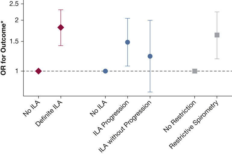
Results: In adjusted models, we found that lower pulmonary vascular volumes on CT were associated with greater odds of ILA, antecedent ILA progression, and restrictive pattern on spirometry. For example, each SD lower small vessel fraction was associated with 1.81-fold greater odds of ILA (95% CI, 1.41-2.31; P < .0001), and 1.63-fold greater odds of restriction on spirometry (95% CI, 1.18-2.24; P = .003). Similar patterns were seen after adjustment for diffusing capacity for carbon monoxide, emphysema, and among participants with normal lung function.
Interpretation: In this cohort of community-dwelling adults not selected on the basis of lung disease, more severe vascular pruning on CT was associated with greater odds of ILA, ILA progression, and restrictive pattern on spirometry. Pruning on CT may be an indicator of early pulmonary vasculopathy associated with interstitial lung disease.
Keywords: epidemiology (pulmonary); imaging; interstitial lung disease; pulmonary circulation.
Copyright © 2020 American College of Chest Physicians. Published by Elsevier Inc. All rights reserved.
Figures

Comment in
-
Vascular Pruning: A Sign of Early Pulmonary Vascular Disease or a Surrogate Marker of Interstitial Lung Abnormalities?Chest. 2021 Feb;159(2):473-474. doi: 10.1016/j.chest.2020.10.006. Chest. 2021. PMID: 33563431 No abstract available.
References
-
- Lettieri C.J., Nathan S.D., Barnett S.D., Ahmad S., Shorr A.F. Prevalence and outcomes of pulmonary arterial hypertension in advanced idiopathic pulmonary fibrosis. Chest. 2006;129(3):746–752. - PubMed
-
- Koschel D.S., Cardoso C., Wiedemann B., Höffken G., Halank M. Pulmonary hypertension in chronic hypersensitivity pneumonitis. Lung. 2012;190(3):295–302. - PubMed
-
- Price L.C., Devaraj A., Wort S.J. Central pulmonary arteries in idiopathic pulmonary fibrosis: size really matters. Eur Respir J. 2016;47(5):1318–1320. - PubMed
Publication types
MeSH terms
Grants and funding
LinkOut - more resources
Full Text Sources
Other Literature Sources
Medical
Research Materials
