. 2020 Nov;88(5):921-932.
doi: 10.1002/ana.25878. Epub 2020 Aug 31.

Sex Mediates Relationships Between Regional Tau Pathology and Cognitive Decline

Affiliations

Sex Mediates Relationships Between Regional Tau Pathology and Cognitive Decline

Rachel F Buckley et al. Ann Neurol. 2020 Nov.

Abstract

Objective: The goal of this study was to examine sex differences in tau distribution across the brain of older adults, using positron emission tomography (PET), and investigate how these differences might associate with cognitive trajectories.

Methods: Participants were 343 clinically normal individuals (women, 58%; 73.8 [8.5] years) and 55 individuals with mild cognitive impairment (MCI; women, 38%; 76.9 [7.3] years) from the Harvard Aging Brain Study and the Alzheimer's Disease Neuroimaging Initiative. We examined 18 F-Flortaucipir (FTP)-positron emission tomography (PET) signal across 41 cortical and subcortical regions of interest (ROIs). Linear regression models estimated the effect of sex on FTP-signal for each ROI after adjusting for age and cohort. We also examined interactions between sex*Aβ-PET positive / negative (+ / -) and sex*apolipoprotein ε4 (APOEε4) status. Linear mixed models estimated the moderating effect of sex on the relationship between a composite of sex-differentiated tau ROIs and cognitive decline.

Results: Women showed significantly higher FTP-signals than men across multiple regions of the cortical mantle (p < 0.007). β-amyloid (Aβ)-moderated sex differences in tau signal were localized to medial and inferio-lateral temporal regions (p < 0.007); Aβ + women exhibited greater FTP-signal than other groups. APOEε4-moderated sex differences in FTP-signal were only found in the lateral occipital lobe. Women with higher FTP-signals in composite ROI exhibited faster cognitive decline than men (p = 0.04).

Interpretation: Tau vulnerability in women is not just limited to the medial temporal lobe and significantly contributed to greater risk of faster cognitive decline. Interactive effects of sex and Aβ were predominantly localized in the temporal lobe, however, sex differences in extra-temporal tau highlights the possibility of accelerated tau proliferation in women with the onset of clinical symptomatology. ANN NEUROL 2020;88:921-932.


Conflict of interest statement

Potential Conflicts of Interest

The authors declared no conflict of interest.

Figures

FIGURE 1:
FIGURE 1:
Main effects of sex on 18F-Flortaucipir (FTP) standardized uptake value ratios (SUVrs) after adjusting for age and cohort (A) with region of interest (ROI)-based analyses across all participants (corrected p < 0.007), (B) showing vertex-wise (false discovery rate [FDR] corrected p = 0.016), and (C) showing beta weights and standard errors for each ROI with corresponding model R2 (bars to the right denoting female > male [F > M]). ns = not significant; PET = positron emission tomography; PVC = partial volume corrected; STS = superior temporal sulcus.
FIGURE 2:
FIGURE 2:
Interactive effects of sex and β-amyloid (Aβ) positive / negative (+ / −)on 18F-Flortaucipir (FTP) standardized uptake value ratios (SUVrs) after adjusting for age and cohort (A) with region of interest (ROI)-based analyses within Aβ + participants only (n = 129; corrected p < 0.007), (B) β standardized and standard error for each ROI with corresponding model R2 (bars to the right denoting female > male [F > M]), and (C) model estimates of FTP SUVrs in the inferior temporal cortex (with 95% confidence intervals) in men and women with Aβ-positron emission tomography (PET) +/−. ns = not significant; PVC = partial volume corrected; STS = superior temporal sulcus.
FIGURE 3:
FIGURE 3:
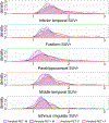
Density maps depicting the distributions of 18F-Flortaucipir (FTP)-signal in regions that are differentiated by a sex*Aβ interaction. Aβ = β-amyloid; PET = positron emission tomography; SUVr = standardized uptake value ratio.
FIGURE 4:
FIGURE 4:
Interactive effects of sex and apolipoprotein ε4 (APOEε4) on 18F-Flortaucipir (FTP) standardized uptake value ratio (SUVr) after adjusting for age and cohort (A) with region of interest (ROI)-based analyses within APOEε4 carriers only (n = 109; corrected p < 0.007), (B) β standardized and standard error for each ROI with corresponding model R2 (bars to the right denoting female > male [F > M]), and (C) model estimates (with 95% confidence interval) of FTP SUVr in men and women with and without APOEε4 carriage. ns = not significant; PET = positron emission tomography; PVC = partial volume corrected; STS = superior temporal sulcus.
FIGURE 5:
FIGURE 5:
Spaghetti plot of cognitive trajectories over time in clinically normal individuals (0th time represents the first 18F-Flortaucipir [FTP]-positron emission tomography [PET] scan) and faceted by a tertile split of composite FTP-PET signal (top = meta-region of interest [ROI], middle = meta temporal ROI, and bottom = reference-ROI) with quadratic fit curves stratified by sex. PACC = Preclinical Alzheimer’s Cognitive Composite.
FIGURE 6:
FIGURE 6:
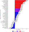
Main effects of sex on 18F-Flortaucipir (FTP) standardized uptake value ratio (SUVr) signal in clinically normal individuals only, showing beta weights and standard errors for each region of interest (ROI) with corresponding model R2 (bars to the right denoting female > male [F > M]). Red indicates multiple comparison significance p < 0.007, purple indicates p < 0.05, and blue indicates subthreshold estimates. PET = positron emission tomography; PVC = partial volume corrected; STS = superior temporal sulcus.
FIGURE 7:
FIGURE 7:
Main effect of sex on 18F-Flortaucipir (FTP) standardized uptake value ratio (SUVr) after adjusting for age and cohort (region of interest [ROI]-based analyses): (A) Alzheimer’s Disease Neuroimaging Initiative (ADNI) uncorrected, (B) ADNI corrected, (C) Harvard Aging Brain Study (HABS) uncorrected, and (D) HABS corrected.

References

    1. Altmann A, Tian L, Henderson VW, Greicius MD. Sex modifies the APOE-related risk of developing Alzheimer disease. Ann Neurol 2014;75:563–573. - PMC - PubMed
    1. Damoiseaux JS, Seeley WW, Zhou J, et a I. Gender modulates the APOE ε4 effect in healthy older adults: convergent evidence from functional brain connectivity and spinal fluid tau levels. J Neurosci 2012;32:8254–8262. - PMC - PubMed
    1. Hohman T, Dumitrescu L, Barnes L, et al. Sex-specific effects of Apolipoprotein E on cerebrospinal fluid levels of tau. JAMA Neurol 2018;75:989–998. - PMC - PubMed
    1. Buckley RF, Mormino EC, Chhatwal J, et al. Associations between baseline amyloid, sex, and APOE on subsequent tau accumulation in cerebrospinal fluid. Neurobiol Aging 2019;78:178–185. - PMC - PubMed
    1. Buckley R, Mormino E, Rabin J, et al. Sex differences in the association between regional tau and global amyloid PET. JAMA Neurol 2019;76:542–551. - PMC - PubMed

Publication types

MeSH terms