. 2021 Mar;288(6):2014-2029.
doi: 10.1111/febs.15524. Epub 2020 Sep 5.

Biochemical characterization of AeD7L2 and its physiological relevance in blood feeding in the dengue mosquito vector, Aedes aegypti

Affiliations

Biochemical characterization of AeD7L2 and its physiological relevance in blood feeding in the dengue mosquito vector, Aedes aegypti

Ines Martin-Martin et al. FEBS J. 2021 Mar.

Abstract

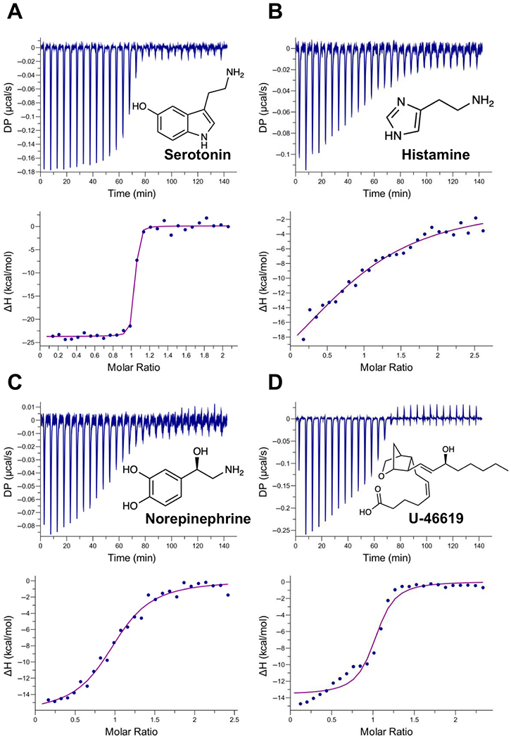
Aedes aegypti saliva facilitates blood meal acquisition through pharmacologically active compounds that prevent host hemostasis. Among these salivary proteins are the D7s, which are highly abundant and have been shown to act as scavengers of biogenic amines and eicosanoids. In this work, we performed comparative structural modeling, characterized the binding capabilities, and assessed the physiological functions of the Ae. aegypti salivary protein AeD7L2 compared to the well-characterized AeD7L1. AeD7L1 and AeD7L2 show different binding affinities to several biogenic amines and biolipids involved in host hemostasis. Interestingly, AeD7L2 tightly binds U-46619, the stable analog of thromboxane A2 (KD = 69.4 nm), which is an important platelet aggregation mediator, while AeD7L1 shows no binding. We tested the ability of these proteins to interfere with the three branches of hemostasis: vasoconstriction, platelet aggregation, and blood coagulation. Pressure myography experiments showed these two proteins reversed isolated resistance artery vasoconstriction induced by either norepinephrine or U-46619. These proteins also inhibited platelet aggregation induced by low doses of collagen or U-46619. However, D7 long proteins did not affect blood coagulation. The different ligand specificity and affinities of AeD7L1 and AeD7L2 matched our experimental observations from studying their effects on vasoconstriction and platelet aggregation, which confirm their role in preventing host hemostasis. This work highlights the complex yet highly specific biological activities of mosquito salivary proteins and serves as another example of the sophisticated biology underlying arthropod blood feeding.

Keywords: arthropods; platelet aggregation; salivary glands; vascular biology; vasodilators.


Conflict of interest statement

Conflict of interest

The authors declare no conflict of interest.

Figures

Fig. 1.
Fig. 1.
Characterization of Aedes aegypti salivary proteins AeD7L1 and AeD7L2. Gene expression analysis of AeD7L1 (A) and AeD7L2 (B) transcripts in different stages of Ae. aegypti mosquitoes. Relative abundance was expressed as the fold change using the 40S ribosomal protein S7 as the housekeeping gene. Larvae stage (reference sample), male pupae, female pupae, and heads and thoraxes (H + T) from male adult and female adult mosquitoes were analyzed separately. Bars represent standard deviation. Two biologicalreplicates were tested. All samples were analyzed in technicaltriplicates. Purification of AeD7L1 (C) and AeD7L2 (D) proteins by size-exclusion chromatography using a Superdex 200 Increase 10/300 GL column. (E) Coomassie-stained NuPAGE Novex 4–12% Bis-Tris gel electrophoresis of the recombinant proteins AeD7L1 and AeD7L2 (5 μg) and Ae. aegypti salivary gland extract (SGE, 10 μg). SeeBlue Plus2 Prestained was used as the protein standard. (F) Western blot showing recognition of recombinant proteins AeD7L1 and AeD7L2 (50 ng) and other protein bands from the salivary gland extract (1.7 μg) by purified IgG recovered from the serum of a rabbit immunized with Ae. aegypti salivary gland extract.
Fig. 2.
Fig. 2.
Binding of AeD7L2 to biogenic amines and U-46619 by isothermaltitration calorimetry. Binding experiments were performed on a VP-ITC microcalorimeter. Assays were performed at 30 °C. The upper curve in each panel shows the measured heat for each injection, while the lower graph shows the enthalpies for each injection and the fit to a single-site binding modelfor calculation of thermodynamic parameters. Panels A–D show binding of AeD7L2 to biogenic amines: serotonin (A), histamine (B), norepinephrine (C), and U-46619 (D). Titration curves are representative of at least two measurements. The insets show the names and chemical formulas for these compounds.
Fig. 3.
Fig. 3.
Sequence alignment of AeD7 long salivary proteins and structure model of AeD7L2. (A) Comparison of Aedes D7 long salivary proteins: AeD7L1 (PDB ID: 3DY9) and AeD7L2 (VectorBase ID: AAEL006417, GenBank ID: AAL16049, or NCBI Reference Sequence: XP_001657778). Sequences without a signal peptide were aligned with Clustal Omega and refined using BoxShade server. Black background shading represents identical amino acids, while gray shading shows similar amino acids. The black arrows indicate predicted amino acids involved in U-46619 binding of AeD7L2 based on the similarity with AnStD7L1 (PDB ID: 3NHT). Red asterisks highlight predicted amino acids involved in serotonin binding for AeD7L2, based on similarity of the model structure with AngaD7R4 (PDB ID: 2QEH). Boxed Tyr-52 is predicted to be involved in TxA2 binding. (B) Superposition of AeD7L1 (PDB ID: 3DY9, shown in gray) and protein structure model of AeD7L2, represented in cyan, shows a similar overall helix structure. (C) Ribbon representation of AeD7L2 protein structure model shows the predicted docking of serotonin in its C-terminal domain binding pocket. (D) Ribbon representation of AeD7L2 protein structure model with U-46619 in its N-terminal domain predicted binding pocket. Amino acid residues of AeD7L2 predicted to be involved in binding are represented as sticks. Serotonin and U-46619 are colored in magenta. The model of the protein structure of AeD7L2 was predicted with I-TASSER software and the structural figures were produced using UCSF Chimera.
Fig. 4.
Fig. 4.
Effect of Aedes aegypti salivary proteins AeD7L1 and AeD7L2 as vasodilators. (A) Experimental setup. Mesenteric arteries isolated from mice were cannulated, pressurized and placed in the myograph chamber. (B) Experimental scheme: Vessel diameter was measured continuously prior to the agonist application (1 μm of NE or U-46619) and for 5 min following the addition of any agonist (baseline), and during and after the addition of 1 μm final concentration of either AeD7L1 or AeD7L2 to the myograph chamber. (C) The inner diameter of mouse isolated mesenteric arteries at baseline (black bar), after the addition of 1 μm of norepinephrine (magenta bar) and following the addition of the recombinant protein AeD7L1 or AeD7L2. (D) The inner diameter of mouse isolated mesenteric arteries at baseline (black bar), after the addition of 1 μm of U-46619 (purple bar) and following the addition of the recombinant protein AeD7L1 or AeD7L2. For each vessel, two measurements of the diameter were averaged (red arrows). For analysis, stable inner diameter values measured during baseline, vasoconstriction, or after the addition of the proteins were used. Results are expressed as the mean of the arterial inner diameter of the biological replicates ± SEM. Results from at least two arteries are represented for each group (four arteries for all experimental groups except when norepinephrine was used as an agonist and AeD7L1 was tested, that only data from two arteries was obtained). Vasoconstriction by NE or U-46619 was indicated as a reduction of diameter taking the baseline value as 100%. Vasodilation achieved by the presence of AeD7L1 or AeD7L2 was expressed as recovery, considering the vasoconstricted diameter as 0% and baseline value as 100%. Snapshots of the myography recordings are shown below each data bar.
Fig. 5.
Fig. 5.
Effect of Aedes aegypti salivary proteins AeD7L1 and AeD7L2 on platelet aggregation. Platelet-rich plasma was preincubated with 1 μm of salivary proteins for 60s before the addition of the platelet aggregation agonists, indicated by an arrow. Platelet aggregation was measured by light transmittance over 6 min. Technical duplicates were run, and SD bars are represented in the figure. (A) Aedes aegypti D7 salivary proteins inhibit collagen-induced platelet aggregation. AeD7L1 shows a delay in platelet shape change and a lower aggregation magnitude. AeD7L2 abolishes collagen-induced platelet aggregation. (B) Platelet aggregation induced by U-46619, the stable analog of Thromboxane A2, was slightly reduced by the presence of AeD7L1 and completely abrogated by AeD7L2. PBS buffer was used as a control (blue). AeD7L1 and AeD7L2 groups are shown in green and magenta, respectively.
Fig. 6.
Fig. 6.
Anticlotting activity of Aedes aegypti salivary gland extracts and AeD7L1 and AeD7L2 salivary proteins. Recalcification time of citrated normal human plasma in the absence (control) or presence of either Ae. aegypti SGE or the recombinant salivary proteins AeD7L1 and AeD7L2. Recalcification or clotting time was determined as the time to reach 0.025 absorption units at 650 nm (onset time to O.D. = 0.025). Exact onset times for Ae. aegypti SGE equivalent to 5 and 2.5 salivary glands could not be determined because coagulation did not occur in these samples during the experimental time of one hour. For plotting and analysis purposes, the maximum experimental time was designated (3600 s). The results are expressed as the mean ± SEM of technical triplicates. Results were analyzed by one-way ANOVA, using the control group as the reference. Statistical differences were set at P < 0.05 (*P < 0.05, ***P < 0.001, ****P < 0.0001).

References

    1. Souza-Neto JA, Powell JR & Bonizzoni M (2019) Aedes aegypti vector competence studies: a review. Infect Genet Evol 67, 191–209. - PMC - PubMed
    1. Ribeiro JM, Rossignol PA & Spielman A (1984) Role of mosquito saliva in blood vessel location. J Exp Biol 108, 1–7. - PubMed
    1. Ribeiro JMC & Arca B (2009) From Sialomes to the Sialoverse: an insight into salivary potion of blood-feeding insects. Adv In Insect Phys 37, 59–118.
    1. Arca B & Ribeiro JM (2018) Saliva of hematophagous insects: a multifaceted toolkit. Curr Opin Insect Sci 29, 102–109. - PubMed
    1. Chagas AC, Ramirez JL, Jasinskiene N, James AA, Ribeiro JM, Marinotti O & Calvo E (2014) Collagen-binding protein, Aegyptin, regulates probing time and blood feeding success in the dengue vector mosquito, Aedes aegypti. Proc Natl Acad Sci U S A 111, 6946–6951. - PMC - PubMed

Publication types

MeSH terms

Substances