Multicenter Study
. 2020 Nov;88(5):1034-1042.
doi: 10.1002/ana.25877. Epub 2020 Sep 9.

Paramagnetic Rim Lesions are Specific to Multiple Sclerosis: An International Multicenter 3T MRI Study

Affiliations
Multicenter Study

Paramagnetic Rim Lesions are Specific to Multiple Sclerosis: An International Multicenter 3T MRI Study

Pietro Maggi et al. Ann Neurol. 2020 Nov.

Abstract

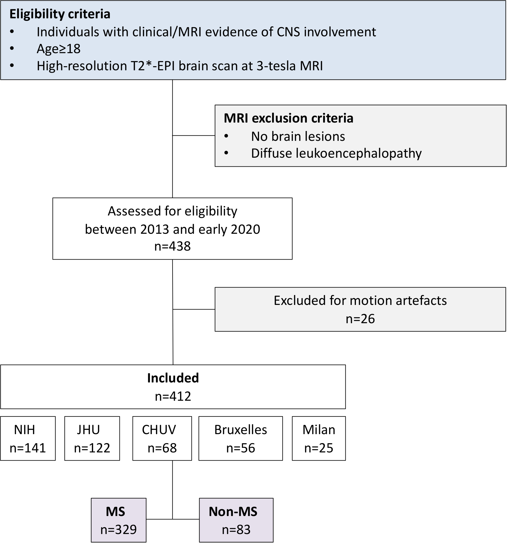
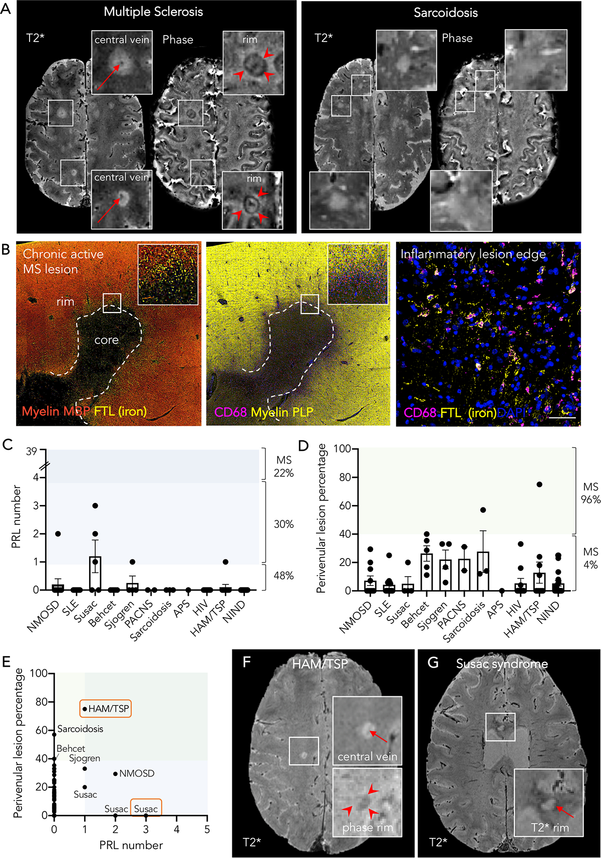
In multiple sclerosis (MS), a subset of chronic active white matter lesions are identifiable on magnetic resonance imaging by their paramagnetic rims, and increasing evidence supports their association with severity of clinical disease. We studied their potential role in differential diagnosis, screening an international multicenter clinical research-based sample of 438 individuals affected by different neurological conditions (MS, other inflammatory, infectious, and non-inflammatory conditions). Paramagnetic rim lesions, rare in other neurological conditions (52% of MS vs 7% of non-MS cases), yielded high specificity (93%) in differentiating MS from non-MS. Future prospective multicenter studies should validate their role as a diagnostic biomarker. ANN NEUROL 2020;88:1034-1042.


Conflict of interest statement

Potential Conflicts of Interest

Nothing to report.

Figures

Figure 1
Figure 1. (A) Typical susceptibility-based MRI features of MS vs non-MS lesions
Representative axial T2* magnitude and phase 3T MR images from individuals with relapsing-remitting multiple sclerosis and sarcoidosis. Whereas MS lesions are generally centered around veins (arrows) and have a hypointense paramagnetic rim (arrowhead), these same features are not typical of non-MS lesions. (B) Representative histopathology of a chronic active rim MS lesion Fluorescent immunostaining of a chronic active lesion in a 58-year old man with secondary progressive MS (31 years of disease; autopsy performed as part of a research program). A dense inflammatory infiltrate of iron-laden (FTL) microglia/macrophages (CD68) characterizes the edge of chronic active/rim lesions in MS and is visible as a paramagnetic rim on susceptibility-based MRI. Chronic active/rim lesions are completely demyelinated without signs of remyelination, as seen with both MBP and PLP myelin staining. Bar scale= 50 μm. (C-G) Frequency of paramagnetic rim lesions and perivenular lesions in non-MS cases (C, D) Distribution of the frequency of PRL (C) and perivenular lesions (D) in the 83 non-MS cases enrolled in the study. Overlapping colored regions refer to the frequency of the same MRI variable in the MS cohort. (E) Identification of cases with at least one PRL and ≥40% perivenular lesions. Only a single case of HAM/TSP satisfied both criteria [MRI images are shown in (F)], and this may be an individual with both diagnoses. (F) Susceptibility-based MRI images of a 49-year-old woman with HAM/TSP, disease duration 10 years, who required two walking aids to walk ~20m without resting. In the inset, 3 of her 4 suprantentorial lesions show some MS-like features including the central vein sign on T2* magnitude images and/or a PRL on phase images. (G) Axial MRI images of a 36-year-old man with Susac disease [cases highlighted in (C)] showing a lesion within the corpus callosum with a rim on T2* images. Abbreviations: MS: multiple sclerosis; FTL: ferritin; MBP: myelin basic protein; PLP: myelin proteolipid protein; PRL: paramagnetic rim lesions; NMOSD: neuromyelitis optica spectrum disorder; SLE: systemic lupus erythematosus; PACNS: primary angiitis of the central nervous system; APS: antiphospholipid syndrome; HIV: human immunodeficiency virus; HAM/TSP: HTLV associated myelopathy/ tropical spastic paraparesis; NIND: non-inflammatory neurological diseases.
Figure 2.
Figure 2.
Flow chart summarizing patients’ progress through the study Site abbreviations: NIH: National Institutes of Health, Clinical Center (Bethesda, MD, USA) JHU: Johns Hopkins University Hospital (Baltimore, MD, USA) CHUV: Lausanne University Hospital (Lausanne, Switzerland) Bruxelles: Erasme University Hospital and Saint Luc University Hospital (Brussels, Belgium) Milan: San Raffaele University Hospital (Milan, Italy)

References

    1. Solomon AJ, Corboy JR. The tension between early diagnosis and misdiagnosis of multiple sclerosis. Nat Rev Neurol 2017;13:567–572. - PubMed
    1. Solomon AJ, Bourdette DN, Cross AH, et al. The contemporary spectrum of multiple sclerosis misdiagnosis: A multicenter study. Neurology 2016;87:1393–1399. - PMC - PubMed
    1. Kaisey M, Solomon AJ, Luu M, Giesser BS, Sicotte NL. Incidence of multiple sclerosis misdiagnosis in referrals to two academic centers. Mult Scler Relat Disord 2019;30:51–56. - PubMed
    1. Tallantyre EC, Dixon JE, Donaldson I, et al. Ultra-high-field imaging distinguishes MS lesions from asymptomatic white matter lesions. Neurology 2011;76:534–539. - PMC - PubMed
    1. Mistry N, Dixon J, Tallantyre E, et al. Central veins in brain lesions visualized with high-field magnetic resonance imaging: a pathologically specific diagnostic biomarker for inflammatory demyelination in the brain. JAMA Neurol 2013;70:623–628. - PubMed

Publication types