

Review
doi: 10.1016/j.disamonth.2020.101062.
Epub 2020 Jul 28.
Severe Acute Respiratory Syndrome Coronavirus (SARS, SARS CoV)
Affiliations
- PMID: 32800504
- PMCID: PMC7386482
- DOI: 10.1016/j.disamonth.2020.101062
Item in Clipboard
Review
Severe Acute Respiratory Syndrome Coronavirus (SARS, SARS CoV)
Dis Mon.
2020 Sep.
No abstract available
Figures
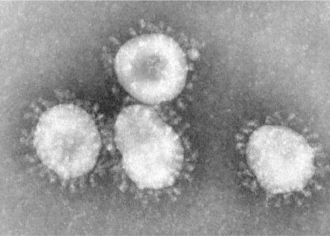
Coronavirus. Centers for Disease Control and Prevention (CDC)/Dr. Fred Murphy.
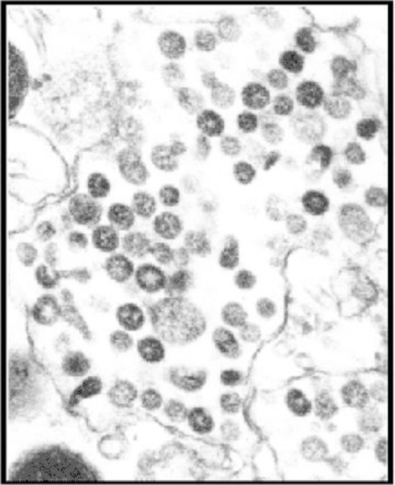
SARS CoV – CDC National Center for Immunization and Respiratory Disease. Division of Viral Diseases.

(Left) CXR SARS Patient – Consider the extensive bilateral ground-glass opacities and poorly defined nodular pattern. In this case diffuse involvement Rt lung, Lt apical sparing. There is mild air-space consolidation is seen in retro-cardiac region of RLL. Mild cardiomegaly present,.

(Right) Bedside supine AP CXR – same patient in Fig. 3, radiograph taken 12 hr after initial radiograph – Note progressive disease in SARS patient, consistent with rapidly declining ARDS. Findings: diffuse bilateral air-space consolidation, prominent air bronchograms. Clinical caveat: note the low position of the endotracheal tube (ETT), and gaseous distention of stomach.,.
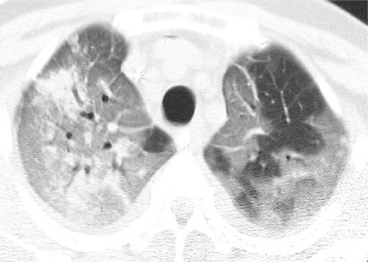
CT Scan Transverse unenhanced image obtained at level of apical segments of upper lobes shows extensive bilateral areas of ground-glass attenuation, more severe on right, and focal areas of consolidation in right upper lobe. Note lobular areas of sparing particularly in left upper lobe,.
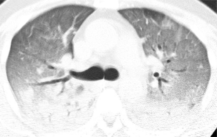
CT image obtained at level of right upper lobe bronchus shows diffuse bilateral areas of ground-glass attenuation and dependent areas of consolidation (37b – 37e).
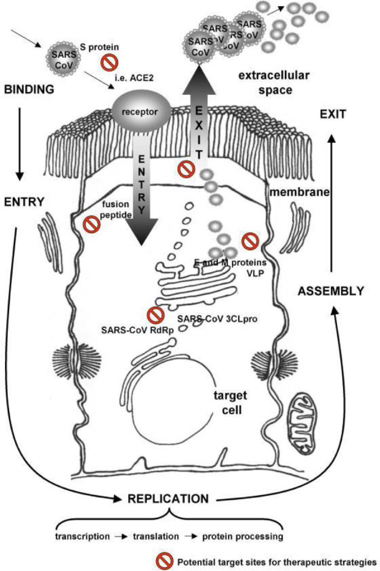
Vaccine research focused on viral structure,
Similar articles
-
COVID-19 - A Brief Review of Radiology Testing.Dis Mon. 2020 Sep;66(9):101059. doi: 10.1016/j.disamonth.2020.101059. Epub 2020 Jul 28. Dis Mon. 2020. PMID: 32912602 Free PMC article. Review. No abstract available.
-
Studies on viral pneumonia related to novel coronavirus SARS-CoV-2, SARS-CoV, and MERS-CoV: a literature review.APMIS. 2020 Jun;128(6):423-432. doi: 10.1111/apm.13047. APMIS. 2020. PMID: 32363707 Review.
-
Current views on the potentials of convalescent plasma therapy (CPT) as Coronavirus disease 2019 (COVID-19) treatment: A systematic review and meta-analysis based on recent studies and previous respiratory pandemics.Rev Med Virol. 2021 Nov;31(6):e2225. doi: 10.1002/rmv.2225. Epub 2021 Feb 23. Rev Med Virol. 2021. PMID: 33621405 Free PMC article.
-
Treatment of COVID-19 with convalescent plasma: lessons from past coronavirus outbreaks.Clin Microbiol Infect. 2020 Oct;26(10):1436-1446. doi: 10.1016/j.cmi.2020.08.005. Epub 2020 Aug 11. Clin Microbiol Infect. 2020. PMID: 32791241 Free PMC article. Review.
-
Introduction - Emerging Pathogens and the COVID-19 Pandemic.Dis Mon. 2020 Sep;66(9):101065. doi: 10.1016/j.disamonth.2020.101065. Epub 2020 Jul 28. Dis Mon. 2020. PMID: 32758362 Free PMC article. Review. No abstract available.
References
-
- Siddell S., Wege H., ter Meulen V. The biology of coronaviruses. J Gen Virol. 1983;64(Pt 4):761–776. - PubMed
-
- Lau S.K.P., Lau C.C.Y., Chan K.H., Li C.P.Y., et al. Delayed induction of proinflammatory cytokines and suppression of innate antiviral response by the novel Middle Easst respiratory syndrome coronavirus: implications for pathogenesis and treatment. J Gen Virol. 2013;94:2679–2690. - PubMed
-
- deGroot R.J., Baker S.C., Baric R., Enjuanes L., et al. Coronaviridae In Virus Taxonomy; Ninth Report of the International Committee On Taxonomy of Viruses pp 806-828. Edited by AMQ King, JH Adams, EB Carstens and EJ Lefkowitz. San Diego, Ca. Elsevier Publishing 2011.
-
- Woo P.C., Lau S.K., Lam C.S., Lau C.C., et al. Disocvery of seven novel mammalian and avian coronaviruses in the genus deltacoronavirus supports bat coronaviruess as the gene source of alphacoronavirus and betacoronavirus and avian cornnaviruses as the gene source of gammacoronavirus and deltacornavirus. J Virol. 2012;86:3995–4008. - PMC - PubMed
Publication types
MeSH terms
Substances
LinkOut - more resources
Full Text Sources
Medical
Miscellaneous
