Review
. 2020 Oct;43(10):780-794.
doi: 10.1016/j.tins.2020.07.004. Epub 2020 Aug 13.

The Distributed Nociceptive System: A Framework for Understanding Pain

Affiliations
Review

The Distributed Nociceptive System: A Framework for Understanding Pain

Robert C Coghill. Trends Neurosci. 2020 Oct.

Abstract

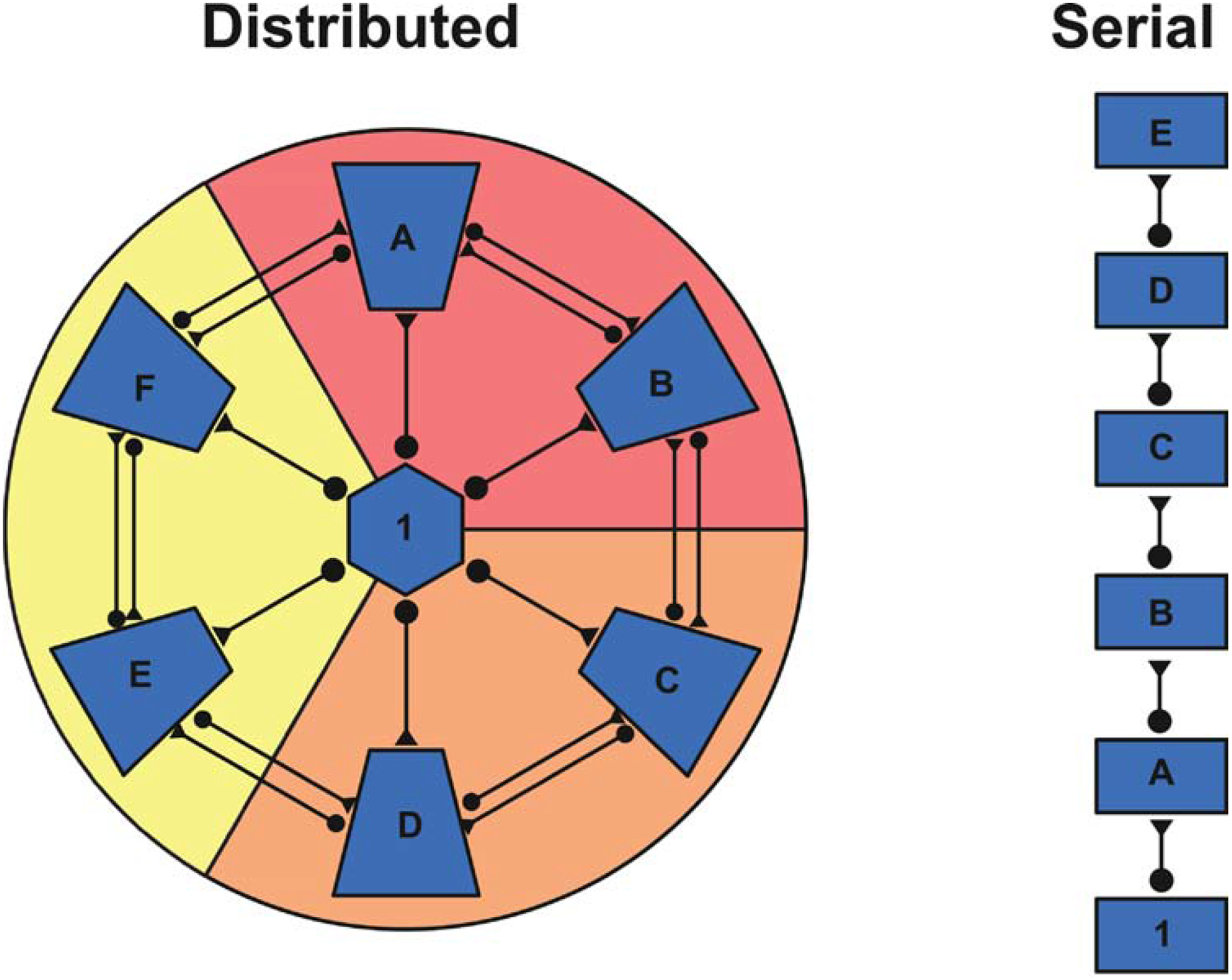
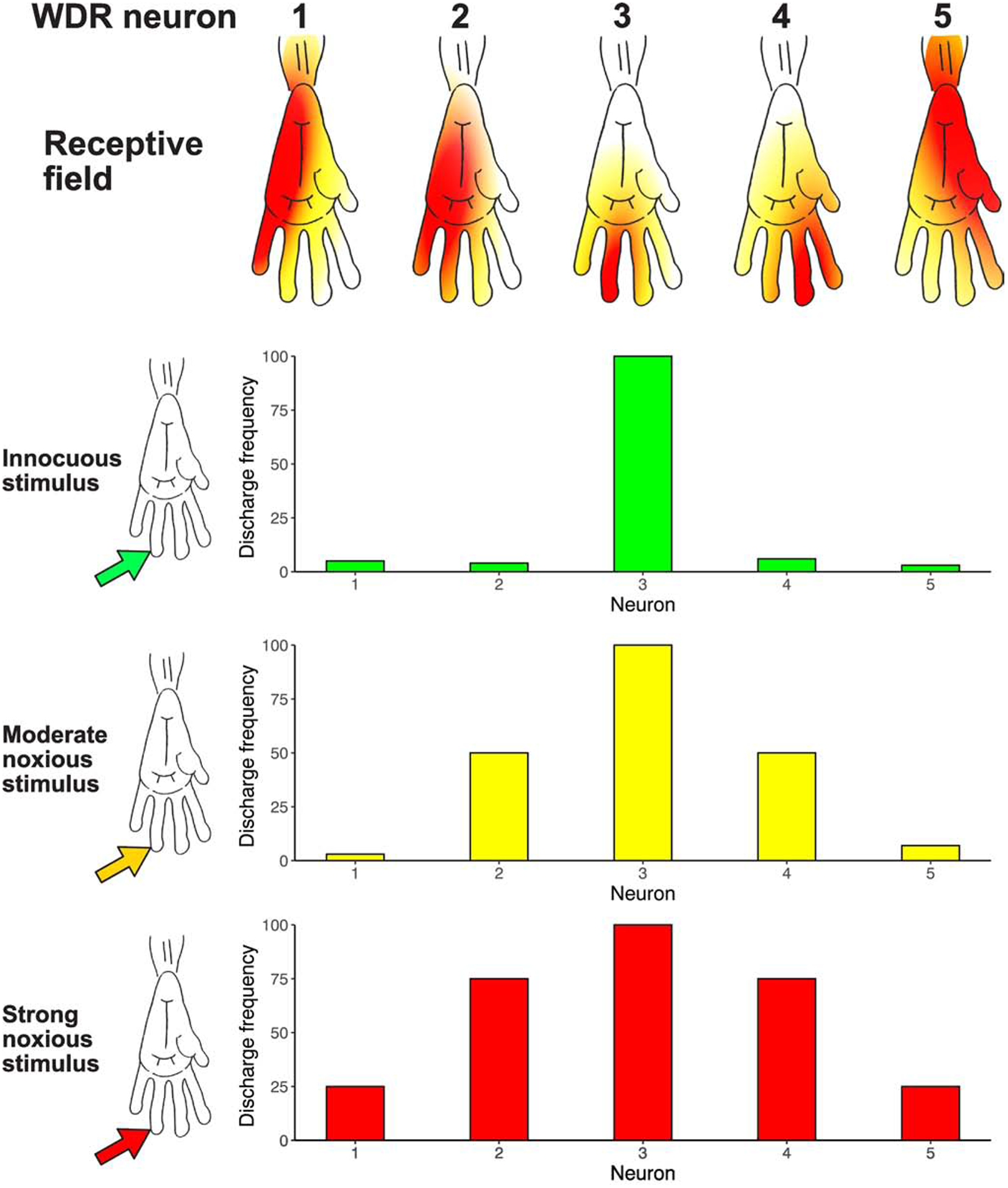
Chronic pain remains challenging to both diagnose and treat. These challenges, in part, arise from limited systems-level understanding of the basic mechanisms that process nociceptive information and ultimately instantiate a subjectively available experience of pain. Here, I provide a framework, the distributed nociceptive system, for understanding nociceptive mechanisms at a systems level by integrating the concepts of neural population coding with distributed processing. Within this framework, wide-spread engagement of populations of neurons produces representations of nociceptive information that are highly resilient to disruption. The distributed nociceptive system provides a foundation for understanding complex spatial aspects of chronic pain and provides an impetus for nonpharmacological cognitive and physical therapies that can effectively target the highly distributed system that gives rise to an experience of pain.

Keywords: bilateral; biomarkers; nociception; pain; population coding; recruitment.


Figures

FIGURE 1
FIGURE 1. Distributed versus Serial Processing of Information
Distributed processing of information can produce a system that is highly resilient to injury whereas serial organizations are significantly more susceptible to disruption. For example, in the distributed system, information flowing from region 1 reaches areas A-F via parallel pathways. In contrast, information flowing from region 1 sequentially needs to pass through all areas prior to reaching area E. Thus, damage to region B would result in minimal disruption of information flow in the distributed organization, while information processing in the serial system would be significantly impacted. Furthermore, a distributed system lends itself to the processing of relatively degenerate information. For example, the modules denoted in red, orange, and yellow could instantiate a representation of information independently of one another. In contrast, serial systems may be crucial when extracting complex features from incoming information.
FIGURE 2
FIGURE 2. Wide Dynamic Range (WDR) Receptive Field Organization: An Architecture for Neural Recruitment and Population Coding
WDR neurons have a complex center/surround receptive field organization [9], and respond to both noxious stimuli as well as light touch in spatially distinct portions of their receptive fields. In this conceptual figure of neurons in the monkey spinal cord, the central receptive field zone (red) of each neuron is responsive to both noxious and innocuous stimuli, while the peripheral surround zone (orange to yellow) is responsive to only noxious stimuli. Moreover, the peripheral surround zone has gradients of sensitivity, such that increasingly intense noxious stimuli are necessary to activate progressively peripheral regions of the surround excitatory receptive field. Given that WDR neurons have relatively large surround receptive fields - frequently encompassing an entire limb or body quadrant - there is likely a substantial overlap of these large surround fields on any given region of the body surface. Accordingly, an innocuous noxious stimulus (green arrow) would recruit a relatively small portion of the population of spinal WDR neurons due to the relatively small receptive field sizes of the central receptive field zone (WDR Neuron #3, red receptive field zone). In contrast, progressively intense noxious stimuli (yellow arrow, red arrow) would be predicted to activate progressively larger portions of the WDR population. Neurons whose central receptive field zones were located within the stimulated area would be activated (WDR Neuron #3), but importantly, neurons whose excitatory surround receptive fields were stimulated would be recruited as well (WDR Neurons #2 and #4 with a moderate noxious stimulus [orange receptive field zone], and then WDR Neurons #1 and 5 with intense noxious stimulus [yellow receptive field zone]). Thus, while single WDR neurons cannot provide sufficient information to distinguish a noxious from an innocuous stimulus, populations of these neurons acting in concert can provide sufficient information to support this distinction [9]. More importantly, noxious stimulus intensity can be encoded by progressive recruitment of increasing numbers of WDR neurons. Portions of this figure are adapted from [82], with permission.
FIGURE 3
FIGURE 3. Spinal Cord Recruitment of Activation in Models of Acute and Chronic Pain
Activation of the rat spinal cord during noxious input was quantified using the 14C-2-deoxyglucose autoradiographic method. This tracer gets taken up by cells within the spinal cord in proportion to glucose utilization, and as such, can provide a marker for neural activity. A. Graded thermal stimuli were applied to the distal hind paw of the rat [17]. As stimulus temperatures increased from neutral (35°C), to mildly noxious (45°C) and to intensely noxious (49°C), graded increases in activity were noted ipsilaterally (right side of the images) within L4, the somatotopic focus of distal hind paw. These increases were located in the superficial dorsal horn, deep dorsal horn, as well as the ventral horn. During intensely noxious stimulation, activation extended far outside of L4, such that significant increases ranged ipsilaterally from L2 through L5, and contralaterally within the deep dorsal horn and ventral horn of L4. This finding confirms predictions of neuron recruitment during graded noxious stimulation inferred from receptive field properties of WDR neurons [9]. B. Vigorous, yet innocuous, brushing of the distal hind paw produced highly focal activation within the intermediate dorsal horn (arrow). This activation was spatially restricted in comparison with the substantial rostral-caudal recruitment of activation evoked by intensely noxious (49°C) stimulation [83]. This finding confirms predictions that noxious stimuli recruit far more neural activity than innocuous stimuli, and further supports the theory that populations of WDR neurons can encode the distinction between noxious and innocuous stimuli [9]. C. Following chronic constriction injury (CCI) of the sciatic nerve - a rat model of neuropathic pain - spinal activation increased dramatically relative to sham operated controls [18]. CCI-related activation extended rostro-caudally from L2 through L5. Moreover, substantial contralateral activation was detected. This extensive ipsilateral and contralateral spread of activity may give rise to the extensive radiation of pain that frequently occurs during complex regional pain syndrome in humans [22]. Modified from [17, 18, 83], with permission.
Figure 4
Figure 4. Schematic Illustration of Anatomic Substrates Supporting Spinal Distribution of Nociceptive Input
There are several anatomical pathways that can support the substantial rostro-caudal distribution of nociceptive activity within the spinal cord. First, when both A-delta and C-fiber afferents enter the spinal cord, they branch considerably in the rostro-caudal direction and traverse relatively long distances within Lissauer’s tract prior to entering the dorsal horn [–86]. These distances are sufficiently large to extend 3 to 7 spinal cord segments (yellow primary afferent denoted by arrow). Accordingly, they can activate ascending neurons within the segment in which they enter (yellow neuron A), as well as neurons several segments rostral or caudal to the segment of entry (orange neuron B). Second, there are substantial propriospinal interconnections that may be sufficient to transmit nociceptive information even further along the rostral-caudal axis of the spinal cord [87, 88], as well as across the midline to the contralateral dorsal horn [89]. These neurons have cell bodies that reside in laminae I, V and VII, and can project more than eight spinal segments (green neuron C). Moreover, they can be activated by noxious stimuli [88]. While typically thought about in the context of motor control, specifically coordinating forelimb with hindlimb activity, they may also provide a substrate for wide ranging facilitation or inhibition of neurons across many spinal segments (red neuron D) [87]. For example, stimuli applied to sites as remote as the forelimb and face can inhibit the responses of noxious stimuli applied to the hindlimb of spinal cord transected monkeys [90]. This integration of sensory input over vast portions of the spinal cord cannot be explained simply by the branching of primary afferents, suggesting that proprio-spinal interconnections are critically involved. Thus, spinal distribution of nociceptive input and potential neuron recruitment may be driven by both widely branching primary afferents as well as by propriospinal interconnections. In addition, this widespread distribution of nociceptive information is key for multi-segmental spatial summation of pain [25].
FIGURE 5. (KEY FIGURE)
FIGURE 5. (KEY FIGURE). Anatomic Substrates for Distribution of Nociceptive Input throughout the Cerebral Cortex
Anatomic studies in primates suggest that nociceptive input may arrive in the cerebral cortex in a highly distributed fashion via parallel routes from the thalamus (middle tier gray shading). Much of this transmission can occur in a di-synaptic fashion, such that a spinal neuron projects directly to a thalamic neuron or other subcortical neuron (yellow), which in turn, projects directly to a cortical neuron (blue) [40]. Cortical regions receiving this parallel, di-synaptic input include the primary somatosensory cortex (SI), secondary somatosensory cortex (SII), the posterior insular cortex, and the anterior cingulate cortex. Of these regions, SI gets input from ventroposterior lateral nucleus (VPL) [91], with subregions 3a,3b,1,and 2 each receiving input [92]; SII gets input from ventroposterior inferior nucleus (VPI) [93]; the granular and dysgranular portions of insula get input from the posterior-suprageniculate (Po-SG) complex [93, 94]; and the cingulate cortex gets input from the mediodorsal nucleus (MD) [95, 96] as well as the parafasicular nucleus [96]; In many instances, this thalamic connectivity is somewhat reciprocal: SI projects to VPL [97], SII projects to VPI [98], the ACC projects to MD [99]. In addition to thalamic routes, subcortical regions such as the globus pallidus (not shown) and central nucleus of the amygdala also receive direct spinal inputs [49], as well as di-synaptic input [40] via non-spinothalamic routes, such as the spinoparabrachial (PB) amygdaloid pathway [100]. This massively parallel architecture provides a clear substrate for direct and widespread distribution of nociceptive information across multiple cerebro-cortical regions. Extensive intra-cortical connectivity augments distribution of afferent nociceptive input within the cerebral cortex. Multiple regions within SI - areas 3a, 3b, 1, and 2- are reciprocally connected with SII [101, 102]. SII is reciprocally connected with the granular and dysgranular portions of the insula [–104]. The granular and dysgranular portions of the insula both project to multiple subregions of the anterior and anterior midcingulate cortex including areas 32, perigenual area 24, as well as 24a, 24b, and 24c (not shown) [105]. In turn, the granular and dysgranular portions of the insula receive inputs from portions of area 24 [103, 106]. The granular insula projects to the lateral nucleus of the amygdala [102], while the dysgranular insula projects to lateral and lateral basal nuclei of the amygdala [102], as well as to the central nucleus of the amygdala [107]. Both granular and dysgranular portions of the insula receive inputs from the basal and basal accessory nuclei of the amygdala [108], while anterior and anterior midcingulate areas 24a and 24b have inputs from the lateral basal and accessory basal nuclei of amygdala [109]. Area 24 also projects to the lateral basal nucleus of the amygdala [106].

References

    1. Neville A et al. (2019) Diagnostic Uncertainty in Youth With Chronic Pain and Their Parents. Journal of Pain 20 (9), 1080–1090. - PubMed
    1. Finnerup NB et al. (2018) Neuropathic pain clinical trials: factors associated with decreases in estimated drug efficacy. Pain 159 (11), 2339–2346. - PMC - PubMed
    1. Price DD et al. (1978) Spatial and temporal transformations of input to spinothalamic tract neurons and their relation to somatic sensations. J Neurophysiol 41 (4), 933–47. - PubMed
    1. Coghill RC et al. (1994) Distributed processing of pain and vibration by the human brain. J Neurosci 14 (7), 4095–108. - PMC - PubMed
    1. Sherrington CS (1908) The integrative action of the nervous system, Archibald Constable.

Publication types