

Review
doi: 10.1016/j.tcb.2020.07.003.
Epub 2020 Aug 13.
EMT, MET, Plasticity, and Tumor Metastasis
Affiliations
- PMID: 32800658
- PMCID: PMC7647095
- DOI: 10.1016/j.tcb.2020.07.003
Item in Clipboard
Review
EMT, MET, Plasticity, and Tumor Metastasis
Trends Cell Biol.
2020 Oct.
Abstract
Cancer cell identity and plasticity are required in transition states, such as epithelial-mesenchymal transition (EMT) and mesenchymal-epithelial transition (MET), in primary tumor initiation, progression, and metastasis. The functional roles of EMT, MET, and the partial state (referred to as pEMT) may vary based on the type of tumor, the state of dissemination, and the degree of metastatic colonization. Herein, we review EMT, MET, pEMT, and plasticity in the context of tumor metastasis.
Keywords: EMT; MET; cellular plasticity; colonization; metastasis; pEMT.
Copyright © 2020 Elsevier Ltd. All rights reserved.
Figures
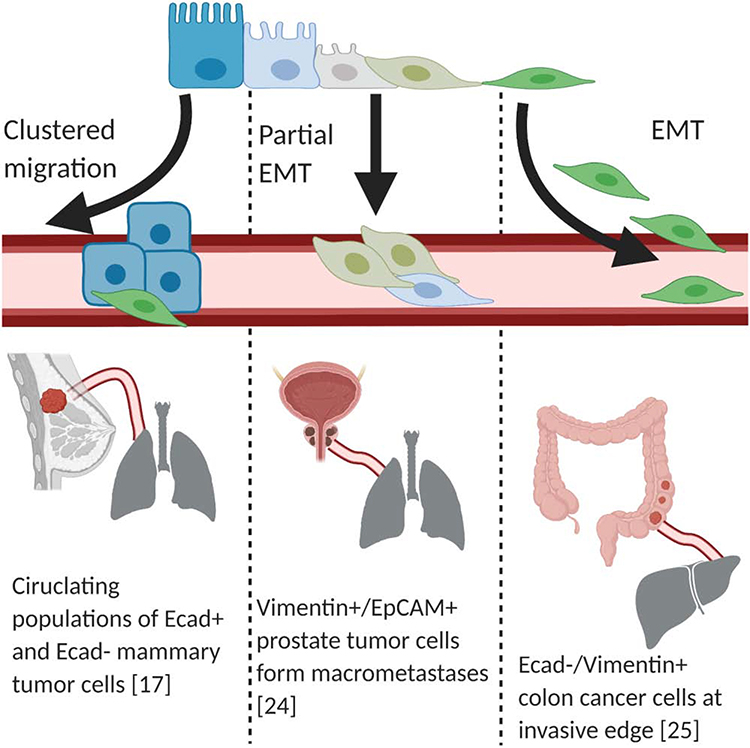
There are multiple mechanisms by which tumor cells (blue rectangles) are thought to detach from the primary site and intravasate into the lymphatic-vascular systems. One proposed mechanism is complete EMT or cEMT (green spheres), whereby cells lose epithelial markers and gain mesenchymal characteristics. During partial EMT or pEMT, cells retain some of their epithelial characteristics. Another possibility is that cells undergoing EMT represent only a small portion of the total population that ultimately metastasize.
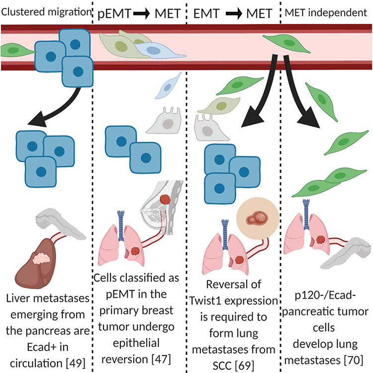
Evidence in multiple model systems demonstrates that macrometastases have strong expression of epithelial markers (blue), which are likely a requirement for expansion within the metastatic site. One model suggests cells that previously underwent cEMT or pEMT regain epithelial markers and lose mesenchymal characteristics (green). Lastly, in mixed populations of metastatic cells, it is thought that the clusters of epithelial cells are the exclusive or dominant population capable of expanding and forming macrometastases.
References
-
- Polyak K and Weinberg RA (2009) Transitions between epithelial and mesenchymal states: Acquisition of malignant and stem cell traits. Nature Reviews Cancer, 9, 265–273 - PubMed
-
- Dongre A and Weinberg RA (2019) New insights into the mechanisms of epithelial–mesenchymal transition and implications for cancer. Nat. Rev. Mol. Cell Biol. 20, 69–84 - PubMed
-
- Pastushenko I and Blanpain C (2019) EMT Transition States during Tumor Progression and Metastasis. Trends in Cell Biology, 29, 212–226 - PubMed
Publication types
MeSH terms
Grants and funding
LinkOut - more resources
Full Text Sources
Medical
Research Materials
Miscellaneous
