. 2020 Oct 1;466(1-2):36-46.
doi: 10.1016/j.ydbio.2020.08.004. Epub 2020 Aug 13.

Pdgfra regulates multipotent cell differentiation towards chondrocytes via inhibiting Wnt9a/beta-catenin pathway during chondrocranial cartilage development

Affiliations

Pdgfra regulates multipotent cell differentiation towards chondrocytes via inhibiting Wnt9a/beta-catenin pathway during chondrocranial cartilage development

Garrett Bartoletti et al. Dev Biol. .

Abstract

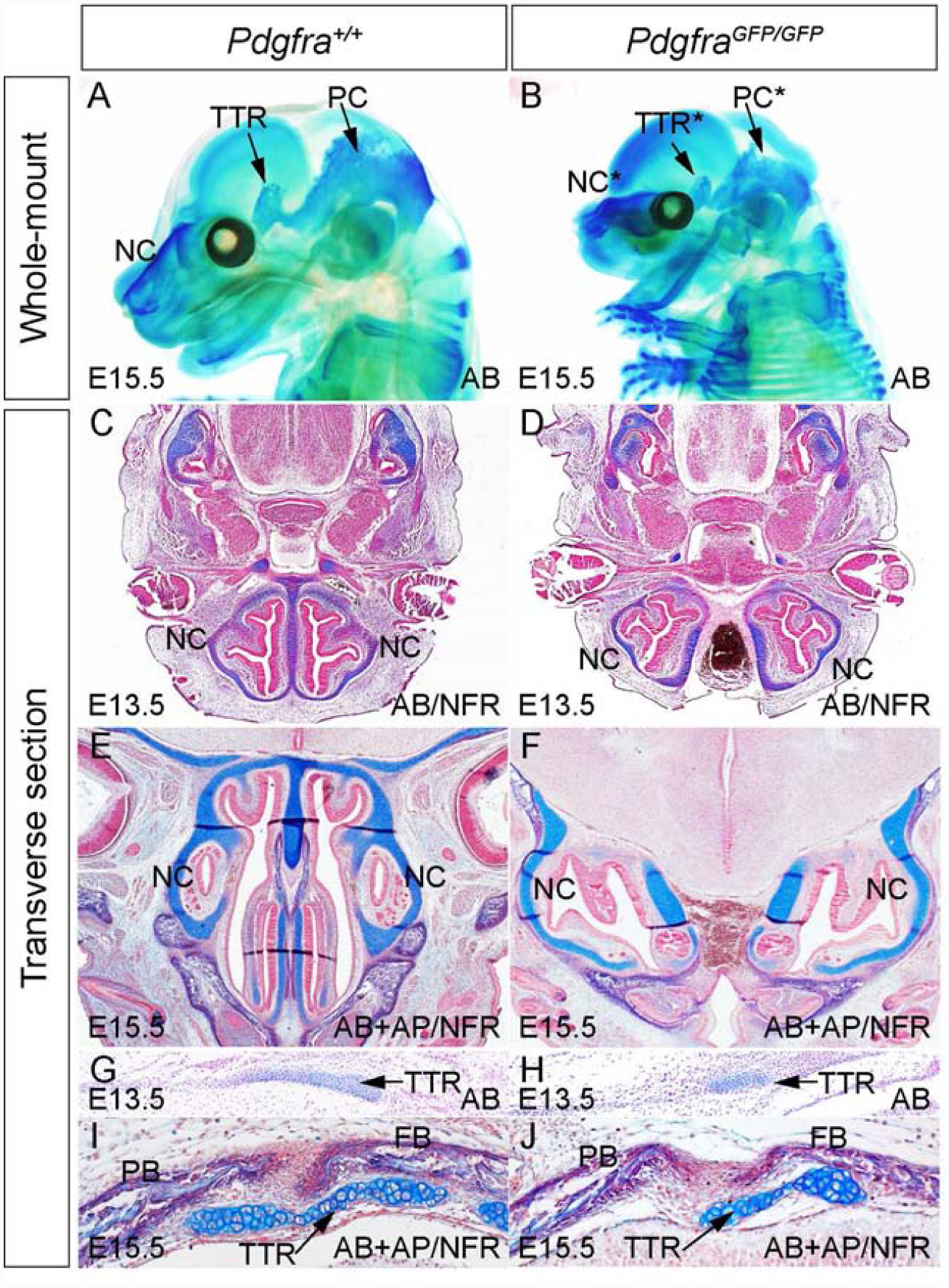
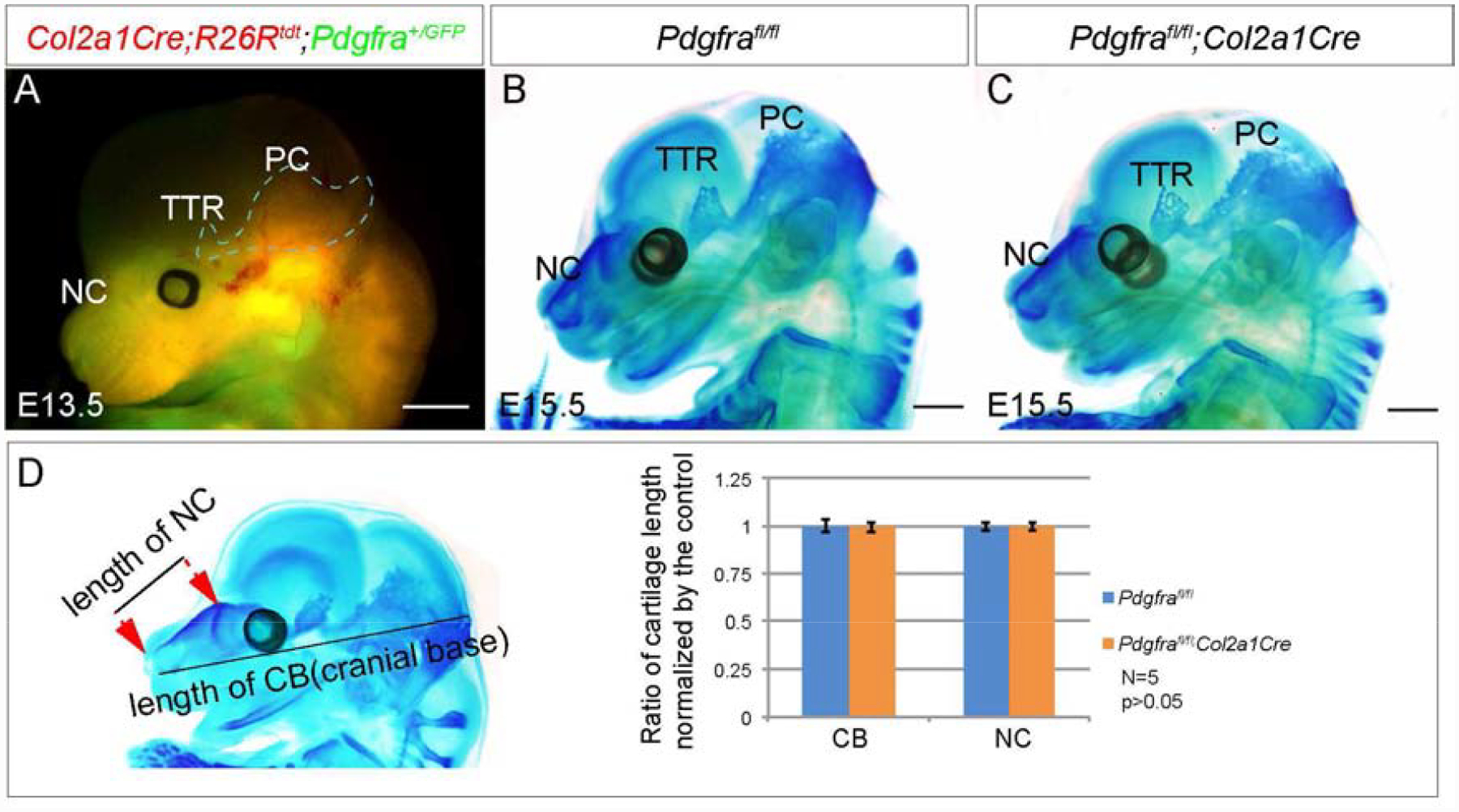
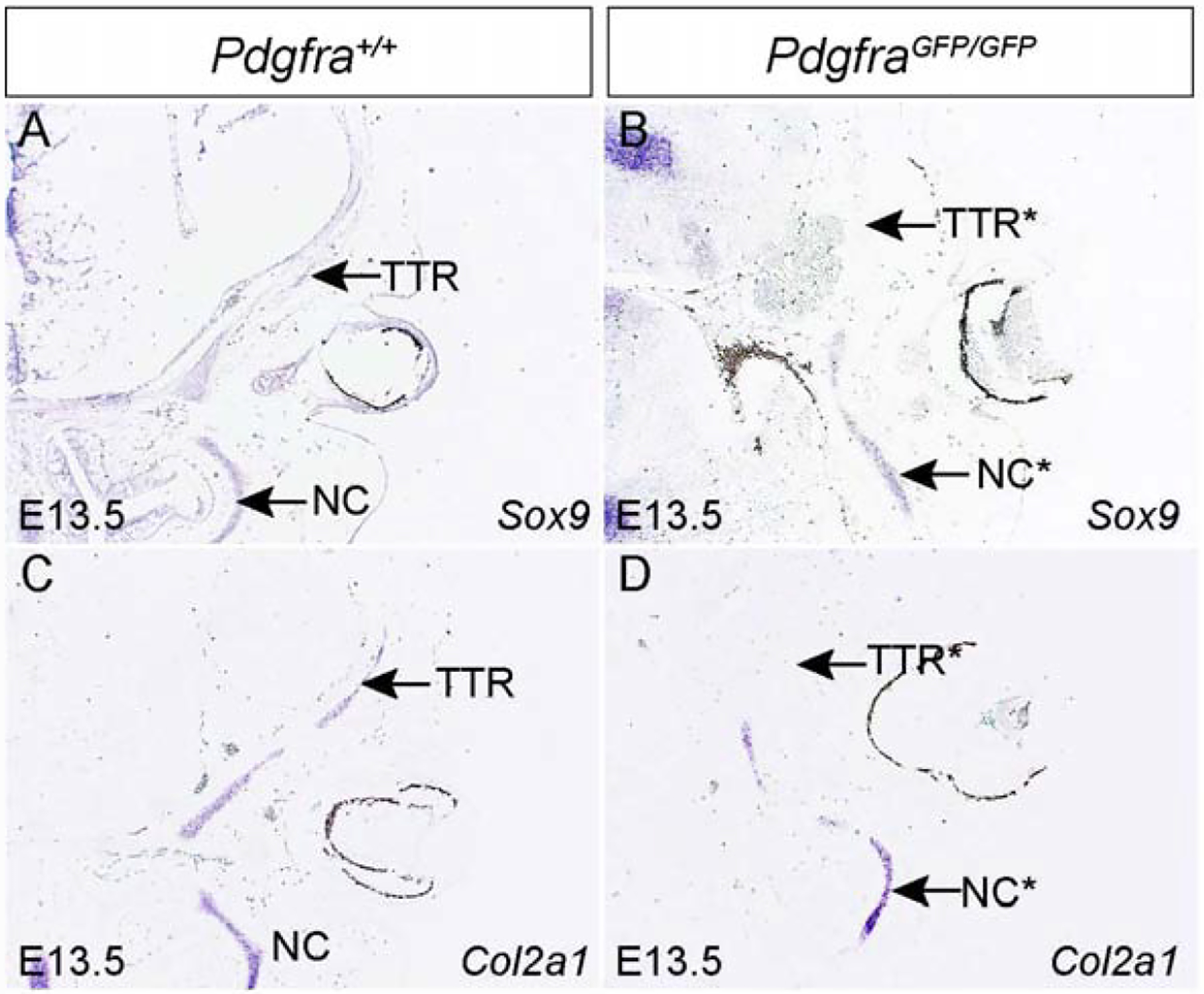
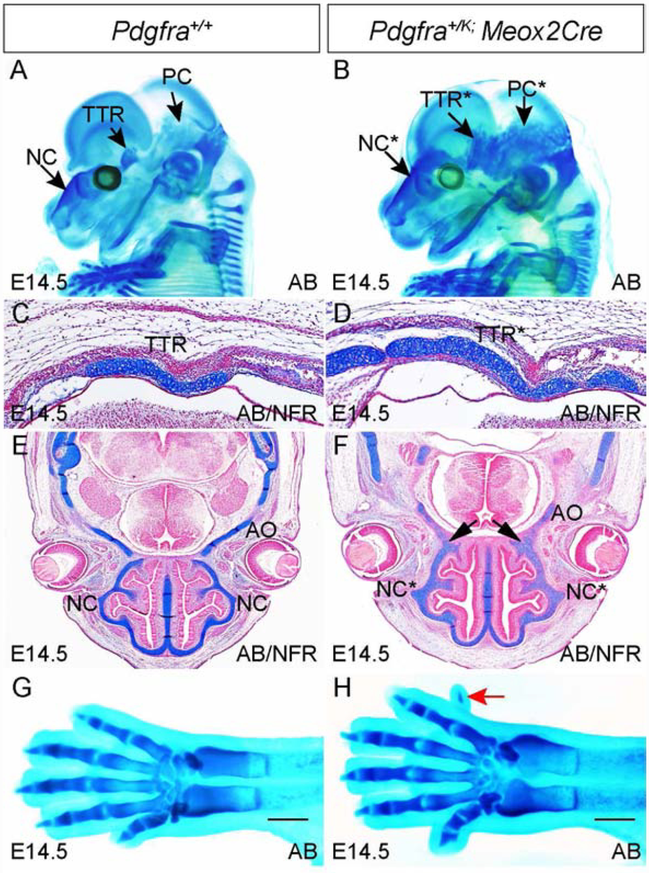
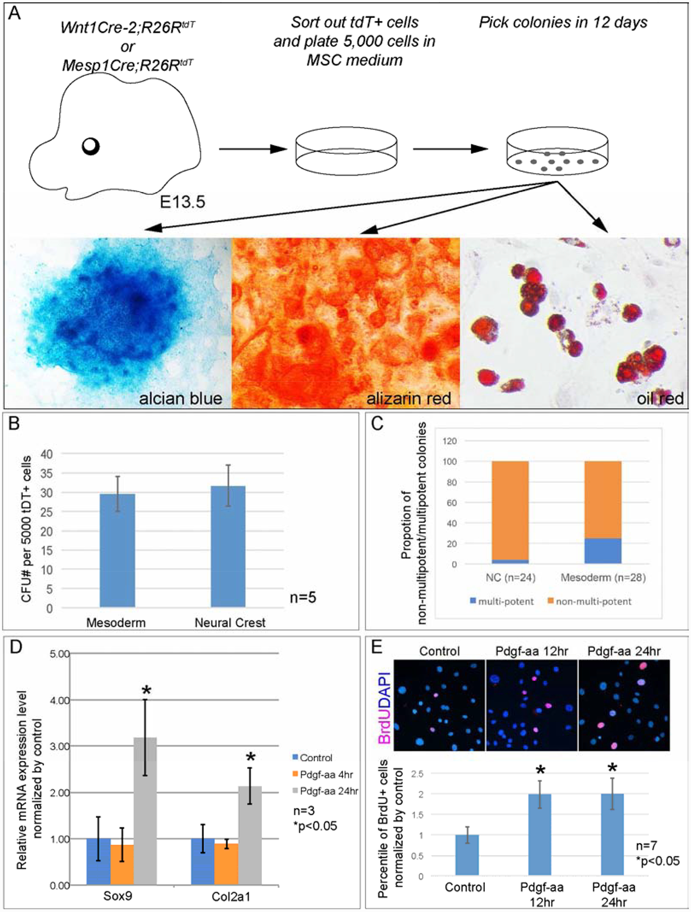
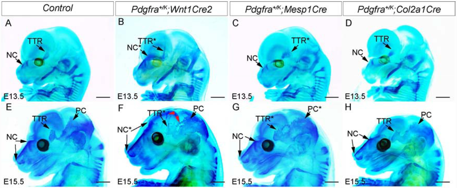
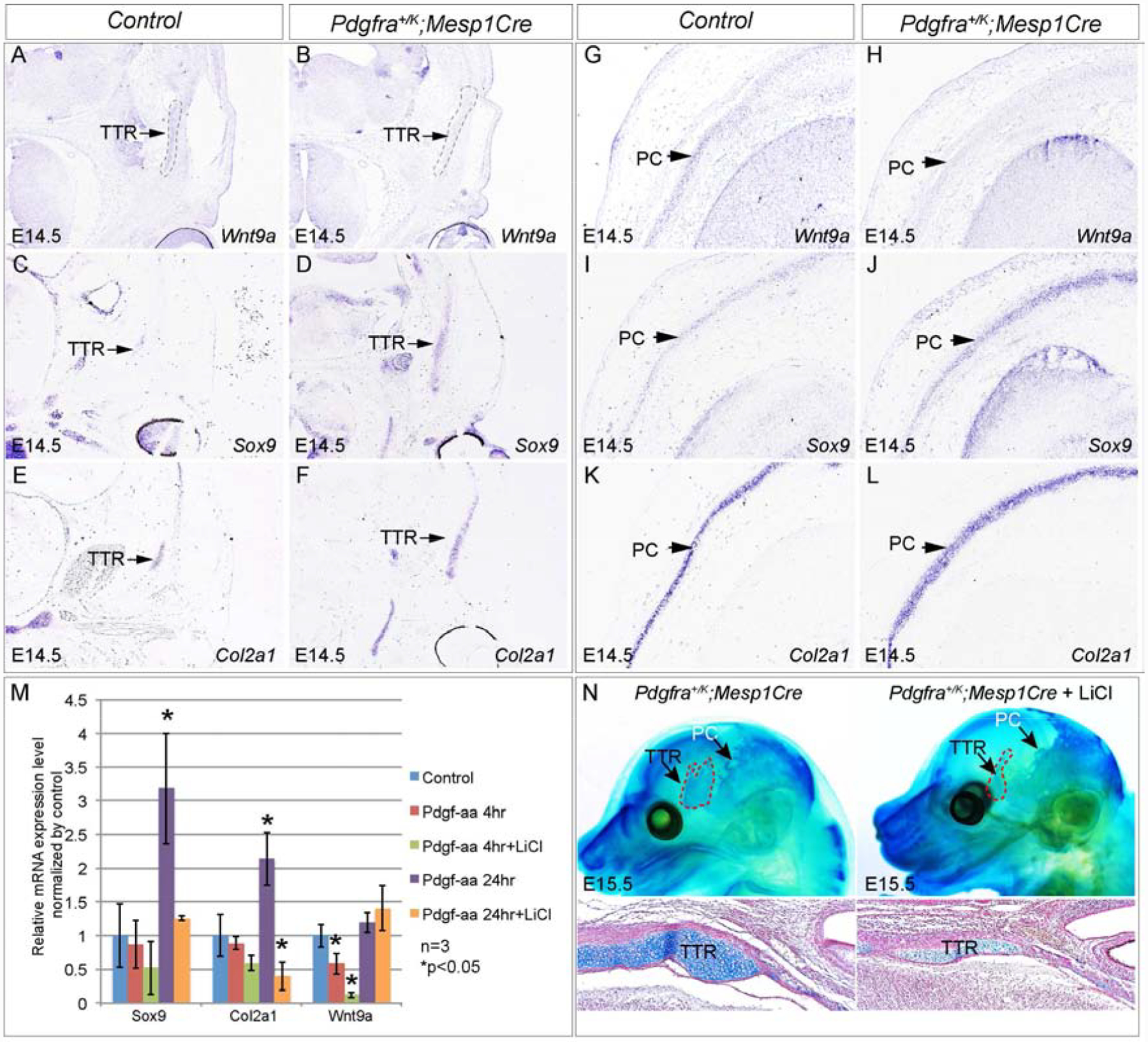
The mammalian skull is composed of the calvarial bones and cartilages. Malformation of craniofacial cartilage has been identified in multiple human syndromes. However, the mechanisms of their development remain largely unknown. In the present study, we identified Pdgfra as a novel player of chondrocranial cartilage development. Our data show that Pdgfra is required for normal chondrocranial cartilage development. Using tissue-specific genetic tools, we demonstrated that Pdgfra is essential for chondrocyte progenitors formation, but not in mature chondrocytes. Further analysis revealed that Pdgfra regulates chondrocytes progenitors development at two stages: in embryonic mesenchymal stem cells (eMSCs), Pdgfra directs their differentiation toward chondrocyte progenitors; in chondrocytes progenitors, Pdgfra activation promotes cell proliferation. We also found that excessive Pdgfra activity causes ectopic cartilage formation. Our data show that Pdgfra directs eMSCs differentiation via inhibiting Wnt9a transcription and its downstream signaling, and activating Wnt signaling rescues ectopic cartilage phenotype caused by excessive Pdgfra activity. In summary, our study dissected the role of Pdgfra signaling in chondrocranial cartilage formation, and illustrated the underlying mechanisms at multiple stages.

Keywords: Chondrocranium; Chondrocyte progenitors; Embryonic mesenchymal stem cells; Pdgfra; Wnt9a.


Conflict of interest statement

Declaration of competing interest The authors declare no competing or financial interests.

Figures

Fig 1.
Fig 1.. Pdgfra is essential for normal chondrocranium development.
(A, B)Whole mount alcian blue staining of Pdgfra+/+ (A) and PdgfraGFP/GFP(B) embryos at E15.5. (C-F) Alcian blue staining on transverse sections across NC of Pdgfra+/+ (C, E) and PdgfraGFP/GFP(D, F) embryos at E13.5(C, D) and at E15.5(E, F). (G-J) Alcian blue staining on transverse sections of Pdgfra+/+ (G, I) and PdgfraGFP/GFP(H, J) embryos at the level of TTR at E13.5(G, H) and at E15.5(I, J). AB, alcian blue; AP, alkaline phosphatase; FB, frontal bone; NC, nasal cartilage; NFR, nuclear fast red; PB, parietal bone; PC, parietal cartilage; TTR, tectum transversum.
Fig 2.
Fig 2.. Pdgfra plays a dispensable role in chondrocyte proliferation and survival.
(A-D) Immunofluorescence staining using anti-cleaved caspase3 antibody(green) on transverse sections of Pdgfra+/+ (A, C) and PdgfraGFP/GFP(B, D) embryos at the level of NC(A, B) and TGG(C, D) at E13.5. The slides were counterstained with DAPI(blue). TGG serves as positive control of the immunostaining assay. (E-H) BrdU labeling on transverse sections of Pdgfra+/+ (E, G) and PdgfraGFP/GFP(F, H) embryos at the level of TTR(E, F) and NC(G, H) at E13.5. (I) Statistical analysis of BrdU labeling results in (E-H). AB, alcian blue; NC, nasal cartilage; NFR, nuclear fast red; TTR, tectum transversum.
Fig 3.
Fig 3.. Chondrocyte-specific inactivation of Pdgfra does not alter chondrocranial cartilage formation.
(A) Co-expression of Pdgfra+ and Col2a1+ cells in E13.5 embryonic head. Pdgfra+ cells are marked with EGFP(green fluorescence) and Col2a1+ cells are marked with tdTomato(red fluorescence) in Col2a1Cre;R26Rtdt;Pdgfra+/GFP embryo. (B, C) Whole mount alcian blue staining of Pdgfrafl/fl(B) and Pdgfrafl/fl;Col2a1Cre(C) embryos at E15.5. (D) Statistical analysis of the length of NC and cranial base in Pdgfrafl/fl and Pdgfrafl/fl;Col2a1Cre embryos at E15.5. N=5, P>0.05. NC, nasal cartilage; PB, parietal bone; PC, parietal cartilage; TTR, tectum transversum. Scale bar= 1mm.
Fig 4.
Fig 4.. Pdgfra deficiency affects chondrocyte progenitor gene expression in the developing chondrocranium.
In situ hybridization results showing chondrocyte progenitor gene Sox9(A, B) and its downstream gene Col2a1(C, D) in Pdgfra+/+ (A, C) and PdgfraGFP/GFP(B, D) embryos at the level of NC and TTR at E13.5. NC, nasal cartilage; TTR, tectum transversum.
Fig 5.
Fig 5.. Augmented Pdgfra activity in epiblasts induces ectopic cartilage formation.
(A, B) Whole mount alcian blue staining of Pdgfra+/+(A) and Pdgfra+/K;Meox2Cre(B) embryos at E14.5. (C-F) Alcian blue staining on transverse sections at the level of TTR(C, D) and NC(E, F) of Pdgfra+/+(C, E) and Pdgfra+/K;Meox2Cre(D, F) embryos at E14.5. Arrows in F point to the fusion between NC and AO. (G, H) Dorsal view of alcian blue staining results of the forelimbs of Pdgfra+/+(G) and Pdgfra+/K;Meox2Cre(H) embryos. Red arrow points to extra digit in H. Scale bar= 1mm. AB, alcian blue; AO, ala
Fig 6.
Fig 6.. Pdgfra activity promotes chondrocyte progenitor formation and proliferation
(A) Procedure of isolating and culturing eMSCs from distinct lineages of E13.5 embryonic heads. (B) Statistical analysis of CFU assay results of primary cells sorted from distinct lineages of E13.5 embryonic craniofacial cells. N=5. (C) Comparison of percentile of multipotent colonies from mesoderm and neural crest cells. (D) Q-PCR result of Sox9 and Col2a1 expression in representative eMSC colony isolated from mesoderm cells. N=3. (E) Statistical analysis of BrdU labeling assay in EGFP+ cells isolated from E12.5 Sox9+/IRES-EGFP embryos. P2 cells are treated with 10ng/ml Pdgf-aa for 12hr or 24hr. Percentile of BrdU+ cells are quantified and compared to untreated cells. N=7.
Fig 7.
Fig 7.. Tissue-specific activation of Pdgfra in chondrocyte progenitors causes ectopic cartilage formation in developing chondrocranium.
(A-H) Whole mount alcian blue staining of the wild-type littermate control(A, E), Pdgfra+/K;Wnt1Cre2(B, F), Pdgfra+/K;Mesp1Cre(C, G) and Pdgfra+/K;Col2a1Cre(D, H) at E13.5 and E15.5. The red arrowheads in F point to ectopic cartilages in Pdgfra+/K;Wnt1Cre2. Tissues with phenotype in mutant mice are marked with asterisks. NC, nasal cartilage; PB, parietal bone; PC, parietal cartilage; TTR, tectum transversum. Scale bar= 1 mm.
Fig 8.
Fig 8.. Wnt9a/beta-catenin signaling mediates Pdgfra regulation on chondrocranium development in vivo.
(A-L) In situ hybridization showing expression of Wnt9a(A, B, G, H), Sox9(C, D, I, J) and Col2a1(E, F, K, L), on transverse sections of the control littermate and Pdgfra+/K;Mesp1Cre embryos at the level of TTR(A-F) and at the level of PC(G-L), respectively. (M) Q-PCR result showing expression of Wnt9a, Sox9 and Col2a1 of a representative eMSC colony treated with Pdgf-aa and LiCl for 4 hours and 24 hours. N=3; asterisk, p<0.05. (N) Whole mount alcian blue staining of Pdgfra+/K;Mesp1Cre and Pdgfra+/K;Mesp1Cre treated with LiCl at E15.5. N=7. Red arrows point to reduction of cartilage formation in LiCl treated embryos. PC, parietal cartilage; TTR, tectum transversum.

References

    1. Akiyama H, Chaboissier MC, Martin JF, Schedl A and de Crombrugghe B (2002). The transcription factor Sox9 has essential roles in successive steps of the chondrocyte differentiation pathway and is required for expression of Sox5 and Sox6. Genes Dev. 16, 2813–2828. - PMC - PubMed
    1. Akiyama H, Lyons JP, Mori-Akiyama Y, Yang X, Zhang R, Zhang Z, Deng JM, Taketo MM, Nakamura T, Behringer RR, et al. (2004). Interactions between Sox9 and beta-catenin control chondrocyte differentiation. Genes Dev. 18, 1072–1087. - PMC - PubMed
    1. Bi W, Deng JM, Zhang Z, Behringer RR and de Crombrugghe B (1999). Sox9 is required for cartilage formation. Nat. Genet 22, 85–89. - PubMed
    1. Brewer JR, Molotkov A, Mazot P, Hoch RV and Soriano P (2015). Fgfr1 regulates development through the combinatorial use of signaling proteins. Genes Dev. 29, 1863–1874. - PMC - PubMed
    1. Captier G, Leboucq N, Bigorre M, Canovas F, Bonnel F, Bonnafe A and Montoya P (2003). Plagiocephaly: morphometry of skull base asymmetry. Surg. Radiol. Anat 25, 226–233. - PubMed

Publication types

MeSH terms

LinkOut - more resources