Review
. 2020 Dec:45:51-64.
doi: 10.1016/j.coviro.2020.07.006. Epub 2020 Aug 13.

Mapping the diverse structural landscape of the flavivirus antibody repertoire

Affiliations
Review

Mapping the diverse structural landscape of the flavivirus antibody repertoire

Madhumati Sevvana et al. Curr Opin Virol. 2020 Dec.

Abstract

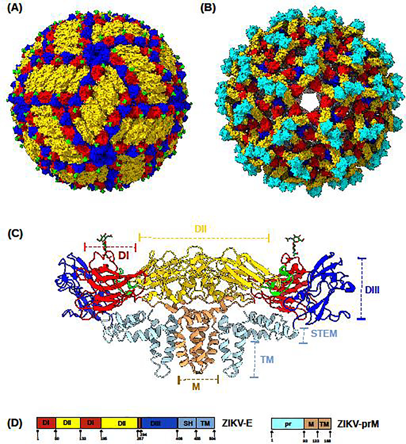
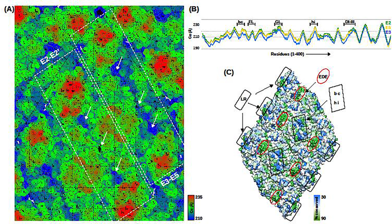
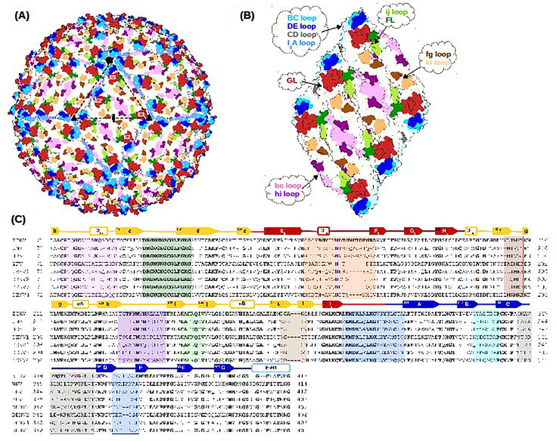
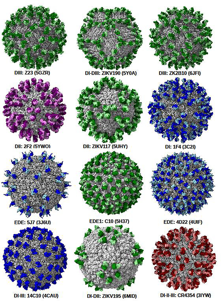
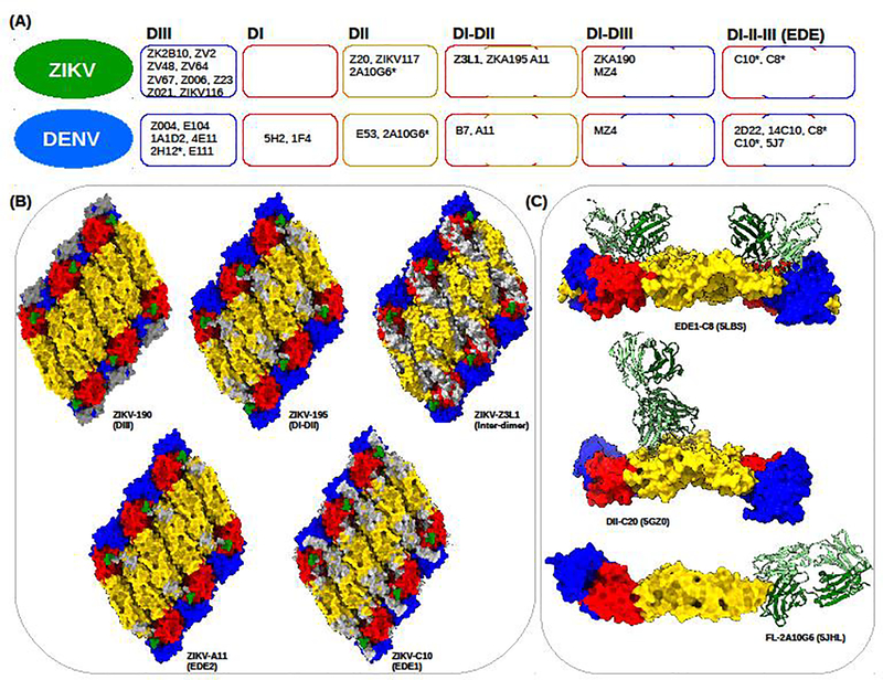
Flaviviruses are emerging arthropod-borne RNA viruses, causing a broad spectrum of life-threatening disease symptoms such as encephalitis and hemorrhagic fever. Successful vaccines exist against yellow fever virus, Japanese encephalitis virus and tick-borne encephalitis virus. However, vaccine development against other flaviviruses like dengue virus is not straightforward. This is partly because of the high sequence conservation and immunological cross-reactivity among flavivirus envelope glycoproteins leading to antibody mediated enhancement of disease. A comprehensive analyses of the structural landscape of humoral immune response against flaviviruses is crucial for antigen design. Here, we compare the available structural data of several flavivirus antibody complexes with a major focus on Zika virus and dengue virus and discuss the mapped epitopes, the stoichiometry of antibody binding and mechanisms of neutralization.


Conflict of interest statement

Conflict of Interest Statement

The authors declare that there is no conflict of interest.

Figures

Figure 1.
Figure 1.. Flavivirus structure
(A) The mature ZIKV structure. (B) The immature ZIKV structure. (C) The secondary structure of flavivirus E and M heterodimer as viewed from the side (parallel to the lipid bilayer). (D) Domain organization of ZIKV structural proteins E and prM/M. In (A), (B), (C) and (D) the domains I (DI), II (DII) and III (DIII) are colored in red, yellow and blue respectively. The glycan is colored in green. The stem helices (SH) and the transmembrane domains (TM) of the E protein are colored light blue. The fusion loop is colored lemon. The membrane protein, M is colored light brown and the pr domain is colored cyan. Cleavage by host protease furin occurs at residue 93 of prM.
Figure 2.
Figure 2.. Non-quasiequivalent icosahedral asymmetric unit.
(A) Landscape of the surface exposed residues in ZIKV colored radially (blue to red) from the center of virus. The two E protein dimers, E2-E2’ and E3-E5 are highlighted in white dashed rectangles. The white arrows point the differences in the surface accessibility of dimer interface residues. (B) Plots showing the distances of the CA distances from the center of the virus. E2, E3 and E5 are colored green, blue and yellow, respectively. (C) Surface rendering of conserved residues on a raft, where the least conserved residues are colored blue and highly conserved residues are colored green. The lateral ridge on DIII (LR), the EDE and the bc and hi loops are highlighted and labelled.
Figure 3.
Figure 3.. The antigenic structure of flaviviruses.
(A) The mature structure of flaviviruses showing the surface exposed antigenic loops of the E proteins. (B) The antigenic loops on an isolated flavivirus raft with each loop labelled. The loops in (A) and (B) are shown in different colors and labelled accordingly. (C) Multiple sequence alignment of flaviviruses. Conserved residues are in bold. The secondary structure elements (SSEs) labelled on top of the ZIKV sequence correspond to ZIKV SSEs. The antigenic loops are highlighted and colored similar to (B).
Figure 4.
Figure 4.. Cryo-EM structures of flavivirus-nFab complexes.
The surface of the virus is colored light grey and rendered viewing along the icosahedral 2-fold axis. Neutralizing Fab complexes of ZIKV, WNV, DENV and JEV are colored green, dark red, blue and magenta, respectively. The nFab is labelled below each structure. The PDB ID corresponding to each structure is shown in parenthesis. The structures are sorted according to their epitope classification.
Figure 5.
Figure 5.. Epitope mapping of representative ZIKV nAbs.
(A) Classification of nAbs against ZIKV and DENV with available 3D structures. (B) Representative ZIKV-Fab epitope mapping (colored white) on ZIKV-raft. The corresponding nAb is labelled under each raft. The targeted domain/s are labelled in parentheses. (C) Three representative crystal structures of ZIKV-Fab complexes: EDE1 targeting C8, DII targeting C20 and FL targeting 2A10G6 are shown. The PDB IDs of the three structures are labelled in parenthesis. In (A), (B) and (C), the domains I, II and III are colored red, yellow and blue respectively. The Fab heavy and light chains are colored green and light green respectively.

Similar articles

Cited by

References

    1. Lindenbach BD, Rice CM: Molecular biology of flaviviruses. Adv. Virus Res 2003, 59:23–61. - PubMed
    1. Guzman MG, Harris E: Dengue. Lancet 2015, 385:453–465. - PubMed
    1. Weaver SC, Costa F, Garcia-Blanco MA, Ko AI, Ribeiro GS, Saade G, Shi PY, Vasilakis N: Zika virus: History, emergence, biology, and prospects for control. Antiviral Res. 2016, 130:69–80. - PMC - PubMed
    1. Butler D: Fears rise over yellow fever’s next move. Nature 2016, 532:155–156. - PubMed
    1. Sejvar JJ: West Nile virus infection. Microbiol Spectr. 2016, 4:EI10–0021-2016. - PubMed

Publication types

MeSH terms

LinkOut - more resources