. 2020 Aug;30(8):1144-1153.
doi: 10.1101/gr.261396.120. Epub 2020 Aug 14.

Detection and characterization of jagged ends of double-stranded DNA in plasma

Affiliations

Detection and characterization of jagged ends of double-stranded DNA in plasma

Peiyong Jiang et al. Genome Res. 2020 Aug.

Abstract

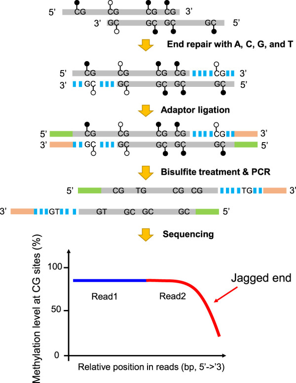
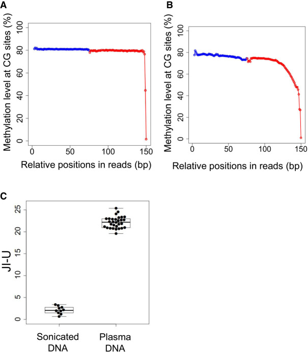
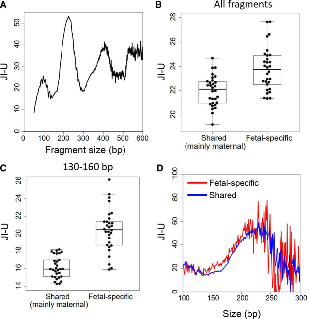
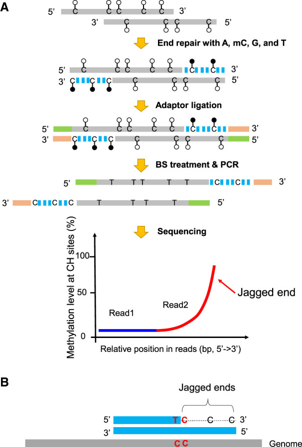
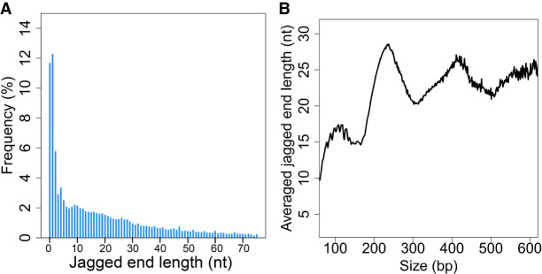
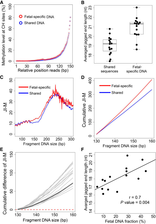
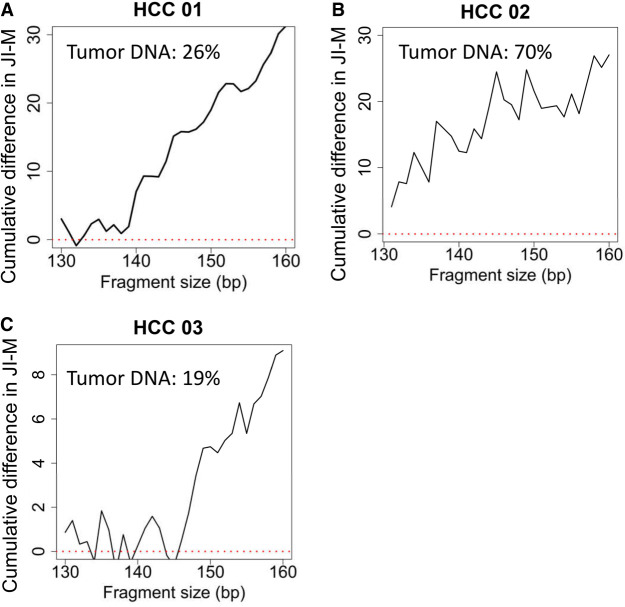
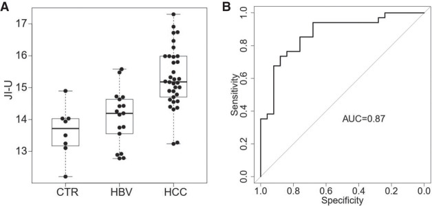
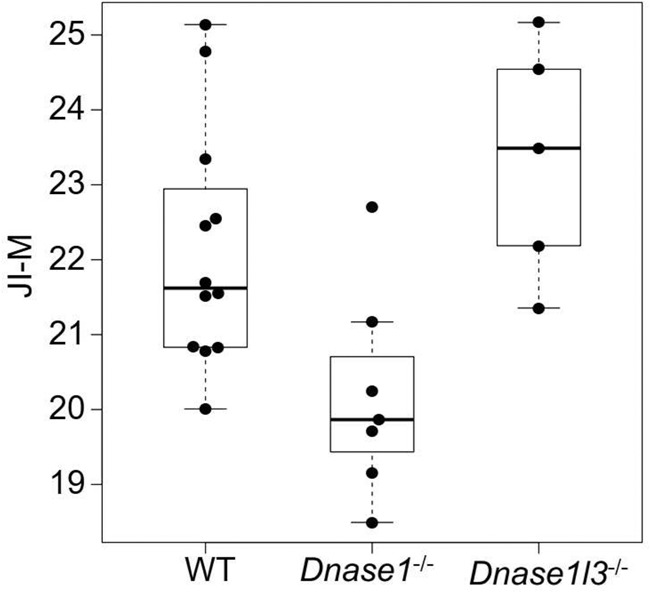
Cell-free DNA in plasma has been used for noninvasive prenatal testing and cancer liquid biopsy. The physical properties of cell-free DNA fragments in plasma, such as fragment sizes and ends, have attracted much recent interest, leading to the emerging field of cell-free DNA fragmentomics. However, one aspect of plasma DNA fragmentomics as to whether double-stranded plasma molecules might carry single-stranded ends, termed a jagged end in this study, remains underexplored. We have developed two approaches for investigating the presence of jagged ends in a plasma DNA pool. These approaches utilized DNA end repair to introduce differential methylation signals between the original sequence and the jagged ends, depending on whether unmethylated or methylated cytosines were used in the DNA end-repair procedure. The majority of plasma DNA molecules (87.8%) were found to bear jagged ends. The jaggedness varied according to plasma DNA fragment sizes and appeared to be in association with nucleosomal patterns. In the plasma of pregnant women, the jaggedness of fetal DNA molecules was higher than that of the maternal counterparts. The jaggedness of plasma DNA correlated with the fetal DNA fraction. Similarly, in the plasma of cancer patients, tumor-derived DNA molecules in patients with hepatocellular carcinoma showed an elevated jaggedness compared with nontumoral DNA. In mouse models, knocking out of the Dnase1 gene reduced jaggedness, whereas knocking out of the Dnase1l3 gene enhanced jaggedness. Hence, plasma DNA jagged ends represent an intrinsic property of plasma DNA and provide a link between nuclease activities and the fragmentation of plasma DNA.


Figures

Figure 1.
Figure 1.
Schematic illustration of plasma DNA jagged end detection using methylation levels at CG sites. A DNA molecule carrying 5′ protruding ends (i.e., jagged ends) would be filled up by the unmethylated nucleotides (i.e., A, C, G, and T) with the presence of T4 DNA polymerase and the Klenow fragment, thus turning into blunt ends. The CG sites present in the newly generated strand were expected to be unmethylated, whereas the CG sites in the original sequence of the same molecule had, on average, a 70% chance of being methylated. The resultant blunted molecules were subjected to bisulfite treatment and PCR amplification. If a molecule contained jagged ends with a 5′ protruding single strand, the methylation levels at CG sites proximal to the 3′ end (e.g., read2) would be lower than that close to the 5′ end (e.g., read1). Filled lollipops represent methylated Cs, and unfilled lollipops represent unmethylated Cs. The dashed blue lines represent newly filled-up nucleotides.
Figure 2.
Figure 2.
Presence of jagged ends in sonicated DNA and plasma DNA. (A) Methylation levels in white blood cell DNA across different CG sites in read1 and read2 after pooling all aligned paired-end reads. The blue dots represent the pooled methylation signal contributed by sequenced CGs at the relative positions in read1. The red dots represent the pooled methylation signal at the relative positions in read2. The methylation signal was calculated by the percentage of sequenced CGs with respect to the total sequenced CGs and TGs. (B) Methylation levels in white blood cell DNA across different CG sites in read1 and read2 after pooling all aligned paired-end reads. (C) JI-U between sonicated DNA and plasma DNA samples. The JI-U represents the jagged index deduced from unmethylated signals at CG sites.
Figure 3.
Figure 3.
Jagged ends in plasma DNA of pregnant women. (A) The profile of JI-U across different sizes of plasma DNA fragments, ranging from 50 bp to 600 bp. (B) The JI-U between shared DNA molecules (mainly of maternal origin) and fetal-specific DNA molecules without size selection. (C) The JI-U between shared DNA molecules and fetal-specific DNA molecules within a range of 130 to 160 bp. (D) JI-U across different sizes for shared and fetal-specific plasma DNA molecules. The JI-U represents the jagged index deduced from unmethylated signals at CG sites.
Figure 4.
Figure 4.
Schematic illustration of plasma DNA jagged end detection using methylation levels at CH sites. (A) The general principle for detecting the presence of jagged ends. In the process of end repair, a DNA molecule carrying 5′ protruding ends (i.e., jagged ends) would be filled up by A, T, mC (methylated C), G with the Klenow fragment without 3′ → 5′ exonuclease activity. The CH (H: A, C, or T) sites present in the newly generated strand were expected to be methylated, whereas the CH sites in the original sequence of the same molecule were generally unmethylated. After the end repair of plasma DNA, the resultant blunted molecules were subjected to bisulfite treatment and PCR amplification. If a molecule contained jagged ends with a 5′ protruding single strand, the methylation levels at CH sites proximal to the 3′ ends (e.g., read2) would be higher than that close to the 5′ end (e.g., read1). Filled lollipops represent methylated Cs, and unfilled lollipops represent unmethylated Cs. The dashed blue lines represent newly filled-up nucleotides. (B) The exact jagged end length deduction. A molecule containing two consecutive cytosines (i.e., “CC” tag) would provide a possibility to directly determine the exact length of a jagged end, when the below criteria are fulfilled: The first one is located within the double-stranded DNA and the other just corresponds to the first nucleotide of the jagged end. Such a “CC” tag would be converted into a “TC” tag after the bisulfite conversion followed by PCR amplification. Therefore, the dashed line indicates the exact jagged end length deduced by CC-tag strategy.
Figure 5.
Figure 5.
The profile of exact jagged end lengths of plasma DNA. (A) Jagged end length distribution deduced by the CC-tag strategy. (B) Averaged jagged end lengths across different sizes of plasma DNA fragments based on CC-tag strategy.
Figure 6.
Figure 6.
Differential jagged ends between fetal and maternal DNA molecules revealed by the incorporation of methylation at CH sites. (A) Methylation level at CH sites in the relative positions of read1 and read2 for shared and fetal-specific DNA molecules. (B) Averaged jagged end lengths between shared and fetal-specific DNA molecules. (C) Relationship between JI-M deduced by methylation at CH sites in read2 and plasma DNA fragment sizes. (D) The cumulative JI-M between fetal-specific and shared DNA molecules. (E) The cumulative difference in JI-M, from 130 bp to 160 bp, between fetal and shared (mainly of maternal origin) DNA sequences. The black line represents the cumulative difference in jaggedness index between the shared (mainly of maternal origin) and fetal-specific DNA molecules pooled from all pregnant subjects. The positive values indicate relative higher jaggedness for molecules below a particular size cutoff. (F) The correlation between the average jagged end length within 130–160 bp and fetal DNA fraction. The JI-M represents the jagged index deduced from the methylated signals at CH sites.
Figure 7.
Figure 7.
The cumulative difference in JI-M between plasma DNA molecules carrying mutant (tumor-derived DNA) and wild-type alleles (mainly nontumoral DNA). The patterns of cumulative difference in JI-M for three plasma DNA samples of patients with HCC, including HCC 01, 02, and 03, are shown in (A), (B), and (C), respectively.
Figure 8.
Figure 8.
Diagnostic performance. (A) Jaggedness indices across healthy control subjects (CTR), patients infected with chronic hepatitis B virus (HBV), and patients with hepatocellular carcinoma (HCC). The y-axis indicates the jaggedness index based on the filling of unmethylated cytosine (JI-U). (B) ROC of jaggedness index between subjects with and without HCC.
Figure 9.
Figure 9.
Jaggedness indices of plasma DNA in mice across different genotypes including wild type, Dnase1−/−, and Dnase1l3−/−. The y-axis indicates the jaggedness index based on the filling of methylated cytosines (JI-M).

Similar articles

Cited by

References

    1. Adams D, Altucci L, Antonarakis SE, Ballesteros J, Beck S, Bird A, Bock C, Boehm B, Campo E, Caricasole A, et al. 2012. BLUEPRINT to decode the epigenetic signature written in blood. Nat Biotechnol 30: 224–226. 10.1038/nbt.2153 - DOI - PubMed
    1. Cabili M, Trapnell C, Goff L, Koziol M, Tazon-Vega B, Regev A, Rinn JL. 2011. Integrative annotation of human large intergenic noncoding RNAs reveals global properties and specific subclasses. Genes Dev 25: 1915–1927. 10.1101/gad.17446611 - DOI - PMC - PubMed
    1. Chan KCA, Zhang J, Hui ABY, Wong N, Lau TK, Leung TN, Lo K-W, Huang DWS, Lo YMD. 2004. Size distributions of maternal and fetal DNA in maternal plasma. Clin Chem 50: 88–92. 10.1373/clinchem.2003.024893 - DOI - PubMed
    1. Chan RWY, Jiang P, Peng X, Tam L-S, Liao GJW, Li EKM, Wong PCH, Sun H, Chan KCA, Chiu RWK, et al. 2014. Plasma DNA aberrations in systemic lupus erythematosus revealed by genomic and methylomic sequencing. Proc Natl Acad Sci 111: E5302–E5311. 10.1073/pnas.1421126111 - DOI - PMC - PubMed
    1. Chan KCA, Jiang P, Sun K, Cheng YKY, Tong YK, Cheng SH, Wong AIC, Hudecova I, Leung TY, Chiu RWK, et al. 2016. Second generation noninvasive fetal genome analysis reveals de novo mutations, single-base parental inheritance, and preferred DNA ends. Proc Natl Acad Sci 113: E8159–E8168. 10.1073/pnas.1615800113 - DOI - PMC - PubMed

Publication types

LinkOut - more resources