Induction of Daptomycin Tolerance in Enterococcus faecalis by Fatty Acid Combinations
- PMID: 32801181
- PMCID: PMC7531955
- DOI: 10.1128/AEM.01178-20
Induction of Daptomycin Tolerance in Enterococcus faecalis by Fatty Acid Combinations
Abstract
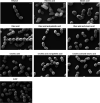
Enterococcus faecalis is a Gram-positive bacterium that normally exists as an intestinal commensal in humans but is also a leading cause of nosocomial infections. Previous work noted that growth supplementation with serum induced tolerance to membrane-damaging agents, including the antibiotic daptomycin. Specific fatty acids found within serum could independently provide tolerance to daptomycin (protective fatty acids), yet some fatty acids found in serum did not and had negative effects on enterococcal physiology (nonprotective fatty acids). Here, we measured a wide array of physiological responses after supplementation with combinations of protective and nonprotective fatty acids to better understand how serum induces daptomycin tolerance. When cells were supplemented with either nonprotective fatty acid, palmitic acid, or stearic acid, there were marked defects in growth and morphology, but these defects were rescued upon supplementation with either protective fatty acid, oleic acid, or linoleic acid. Membrane fluidity decreased with growth in either palmitic or stearic acid alone but returned to basal levels when a protective fatty acid was supplied. Daptomycin tolerance could be induced if a protective fatty acid was provided with a nonprotective fatty acid, and some specific combinations protected as well as serum supplementation. While cell envelope charge has been associated with tolerance to daptomycin in other Gram-positive bacteria, we concluded that it does not correlate with the fatty acid-induced protection we observed. Based on these observations, we conclude that daptomycin tolerance by serum is driven by specific, protective fatty acids found within the fluid.IMPORTANCE With an increasing prevalence of antibiotic resistance in the clinic, we strive to understand more about microbial defensive mechanisms. A nongenetic tolerance to the antibiotic daptomycin was discovered in Enterococcus faecalis that results in the increased survival of bacterial populations after treatment with the drug. This tolerance mechanism likely synergizes with antibiotic resistance in the clinic. Given that this tolerance phenotype is induced by incorporation of fatty acids present in the host, it can be assumed that infections by this organism require a higher dose of antibiotic for successful eradication. The mixture of fatty acids in human fluids is quite diverse, with little understanding between the interplay of fatty acid combinations and the tolerance phenotype we observe. It is crucial to understand the effects of fatty acid combinations on E. faecalis physiology if we are to suppress the tolerance physiology in the clinic.
Keywords: Enterococcus faecalis; daptomycin; fatty acid; membrane fluidity.
Copyright © 2020 Brewer et al.
Figures


Similar articles
-
Exogenous Fatty Acids Protect Enterococcus faecalis from Daptomycin-Induced Membrane Stress Independently of the Response Regulator LiaR.Appl Environ Microbiol. 2016 Jun 30;82(14):4410-4420. doi: 10.1128/AEM.00933-16. Print 2016 Jul 15. Appl Environ Microbiol. 2016. PMID: 27208105 Free PMC article.
-
Incorporation of exogenous fatty acids protects Enterococcus faecalis from membrane-damaging agents.Appl Environ Microbiol. 2014 Oct;80(20):6527-38. doi: 10.1128/AEM.02044-14. Epub 2014 Aug 15. Appl Environ Microbiol. 2014. PMID: 25128342 Free PMC article.
-
Enterococcus faecalis Responds to Individual Exogenous Fatty Acids Independently of Their Degree of Saturation or Chain Length.Appl Environ Microbiol. 2017 Dec 15;84(1):e01633-17. doi: 10.1128/AEM.01633-17. Print 2018 Jan 1. Appl Environ Microbiol. 2017. PMID: 29079613 Free PMC article.
-
In vitro activity of daptomycin against clinical isolates with reduced susceptibilities to linezolid and quinupristin/dalfopristin.Int J Antimicrob Agents. 2006 Nov;28(5):385-8. doi: 10.1016/j.ijantimicag.2006.07.017. Int J Antimicrob Agents. 2006. PMID: 17046205 Review.
-
Daptomycin: a new drug class for the treatment of Gram-positive infections.Drugs Today (Barc). 2005 Feb;41(2):81-90. doi: 10.1358/dot.2005.41.2.882660. Drugs Today (Barc). 2005. PMID: 15821781 Review.
Cited by
-
Emerging mechanisms by which endocannabinoids and their derivatives modulate bacterial populations within the gut microbiome.Adv Drug Alcohol Res. 2023 Dec 8;3:11359. doi: 10.3389/adar.2023.11359. eCollection 2023. Adv Drug Alcohol Res. 2023. PMID: 38389811 Free PMC article. Review.
-
Bacterial cell membranes and their role in daptomycin resistance: A review.Front Mol Biosci. 2022 Nov 14;9:1035574. doi: 10.3389/fmolb.2022.1035574. eCollection 2022. Front Mol Biosci. 2022. PMID: 36452455 Free PMC article. Review.
-
Dual stable isotopes enhance lipidomic studies in bacterial model organism Enterococcus faecalis.Anal Bioanal Chem. 2023 Jul;415(17):3593-3605. doi: 10.1007/s00216-023-04750-3. Epub 2023 May 19. Anal Bioanal Chem. 2023. PMID: 37204445
-
Unexpected contribution of the Fak system and the thioesterase TesE to the growth and membrane physiology of Enterococcus faecalis.J Bacteriol. 2025 Jul 24;207(7):e0012125. doi: 10.1128/jb.00121-25. Epub 2025 Jun 30. J Bacteriol. 2025. PMID: 40586593 Free PMC article.
-
Enterococcus faecalis Readily Adapts Membrane Phospholipid Composition to Environmental and Genetic Perturbation.Front Microbiol. 2021 May 21;12:616045. doi: 10.3389/fmicb.2021.616045. eCollection 2021. Front Microbiol. 2021. PMID: 34093456 Free PMC article.
References
-
- Garsin DA, Frank KL, Silanpaa J, Ausubel FM, Hartke A, Shankar N, Murray BE. 2014. Pathogenesis and models of enterococcal infection In Gilmore MS, Clewell DB, Ike Y, Shankar N (ed), Enterococci: from commensals to leading causes of drug resistant infection. Massachusetts Eye and Ear Infirmary, Boston, MA: https://www.ncbi.nlm.nih.gov/books/NBK190424/. - PubMed
-
- Kristich CJ, Rice LB, Arias CA. 2014. Enterococcal infection-treatment and antibiotic resistance In Gilmore MS, Clewell DB, Ike Y, Shankar N (ed), Enterococci: from commensals to leading causes of drug resistant infection. Massachusetts Eye and Ear Infirmary, Boston, MA: https://www.ncbi.nlm.nih.gov/books/NBK190424/. - PubMed
Publication types
MeSH terms
Substances
Grants and funding
LinkOut - more resources
Full Text Sources
Medical

