. 2020 Sep 1;117(35):21576-21587.
doi: 10.1073/pnas.2007412117. Epub 2020 Aug 14.

Resolution of eicosanoid/cytokine storm prevents carcinogen and inflammation-initiated hepatocellular cancer progression

Affiliations

Resolution of eicosanoid/cytokine storm prevents carcinogen and inflammation-initiated hepatocellular cancer progression

Anna Fishbein et al. Proc Natl Acad Sci U S A. .

Abstract

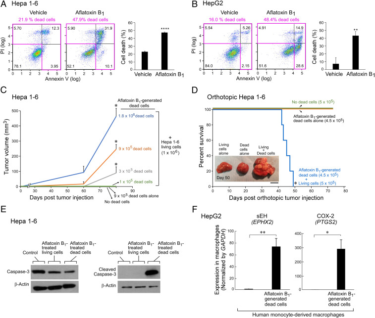
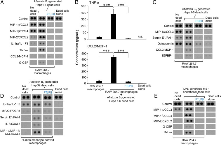
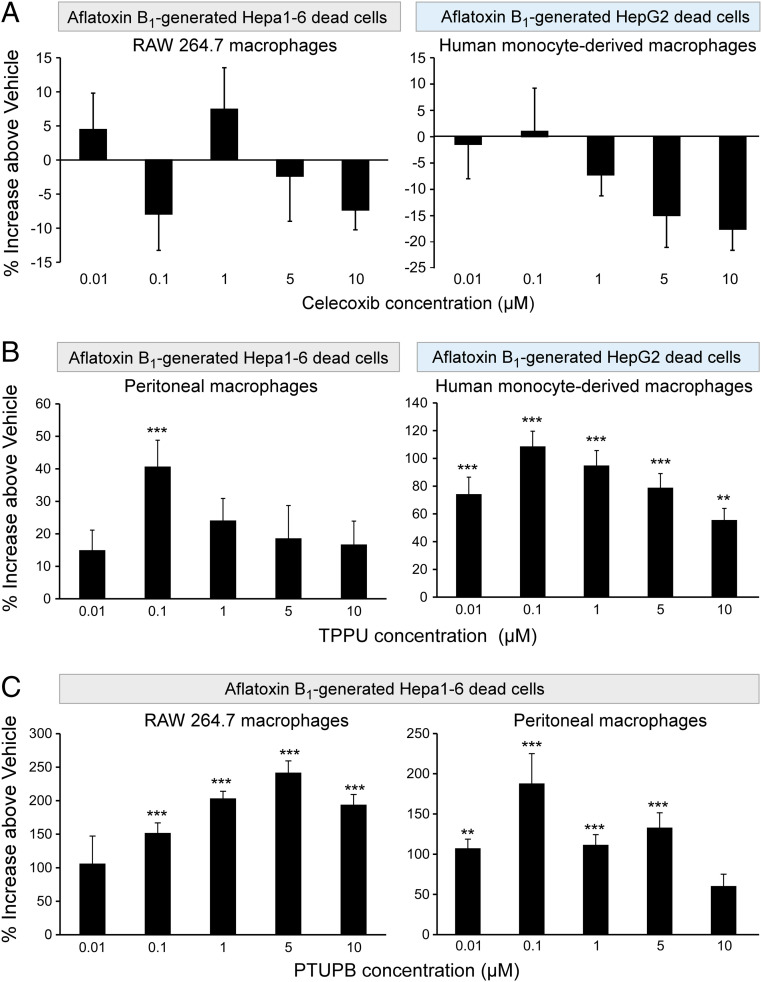
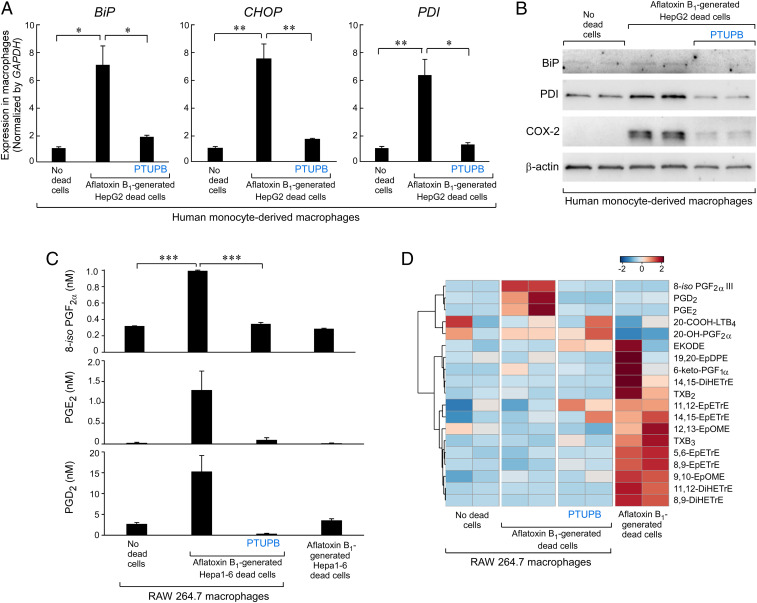
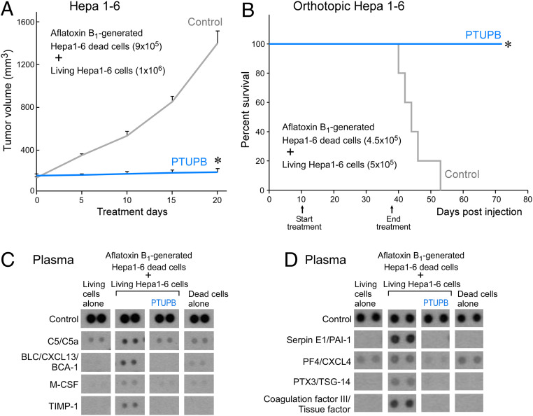
Toxic environmental carcinogens promote cancer via genotoxic and nongenotoxic pathways, but nongenetic mechanisms remain poorly characterized. Carcinogen-induced apoptosis may trigger escape from dormancy of microtumors by interfering with inflammation resolution and triggering an endoplasmic reticulum (ER) stress response. While eicosanoid and cytokine storms are well-characterized in infection and inflammation, they are poorly characterized in cancer. Here, we demonstrate that carcinogens, such as aflatoxin B1 (AFB1), induce apoptotic cell death and the resulting cell debris stimulates hepatocellular carcinoma (HCC) tumor growth via an "eicosanoid and cytokine storm." AFB1-generated debris up-regulates cyclooxygenase-2 (COX-2), soluble epoxide hydrolase (sEH), ER stress-response genes including BiP, CHOP, and PDI in macrophages. Thus, selective cytokine or eicosanoid blockade is unlikely to prevent carcinogen-induced cancer progression. Pharmacological abrogation of both the COX-2 and sEH pathways by PTUPB prevented the debris-stimulated eicosanoid and cytokine storm, down-regulated ER stress genes, and promoted macrophage phagocytosis of debris, resulting in suppression of HCC tumor growth. Thus, inflammation resolution via dual COX-2/sEH inhibition is an approach to prevent carcinogen-induced cancer.

Keywords: bioactive lipid; carcinogenesis; cell death; inflammation resolution; soluble epoxide hydrolase.


Conflict of interest statement

The authors declare no competing interest.

Figures

Fig. 1.
Fig. 1.
AFB1-generated cell death stimulates HCC via up-regulation of sEH and COX-2. Annexin V/PI analysis of (A) Hepa 1-6 murine and (B) HepG2 human HCC tumor cells treated with AFB1 (25 µM, 48 h) compared to vehicle. Quadrants containing dead tumor cells are outlined in pink. Bar graphs represent means of percent cell death ±SEM. n = 3 per group. **P < 0.01, ****P < 0.0001 vs. vehicle. (C) Hepa 1-6 tumor growth stimulated by AFB1-generated dead cells coinjected with a subthreshold inoculum of living cells. n = 5 to 15 mice per group. The two-tailed unpaired Student’s t test was used for final tumor measurements throughout. *P < 0.05 vs. 106 living cells alone (no dead cells). (D) Percent survival of mice coinjected orthotopically into the liver with AFB1-generated Hepa 1-6 dead cells and a subthreshold inoculum of Hepa 1-6 living cells. n = 5 mice per group. Kaplan–Meier analysis indicated significantly shortened survival of mice injected with a combination of dead and living cells as depicted by the area under the Kaplan–Meier survival curves. *P < 0.05. (Scale bar, 1 cm.) (E) Western blot analysis of caspase-3 and cleaved caspase-3 expression in killed and surviving Hepa 1-6 tumor cells treated with AFB1 (25 µM, 48 h) or vehicle. Control = vehicle-treated Hepa 1-6 living cells. Levels of β-actin demonstrate protein loading. (F) Gene expression of sEH (EPHX2) and COX-2 (PTGS2) in human monocyte-derived macrophages coincubated with AFB1-generated HepG2 dead cells; analyzed by qPCR and normalized by GAPDH. n = 3 per group. *P < 0.05, **P < 0.01 vs. control.
Fig. 2.
Fig. 2.
Cytokine storm triggered by carcinogen debris-stimulated macrophages is prevented by dual eicosanoid pathway inhibition. (AC) Inflammatory and angiogenic cytokine array of conditioned media from RAW 264.7 murine macrophage treated with vehicle or PTUPB (5 µM, 2 h) and stimulated with AFB1-generated Hepa 1-6 tumor cell debris vs. debris alone without macrophages. ELISA quantification of TNF-⍺ and CCL2/MCP-1 released by RAW 264.7 macrophages treated with vehicle or PTUPB (5 µM, 2 h) and stimulated with AFB1-generated Hepa 1-6 tumor cell debris. Data are presented as means (pg/mL) ± SEM. n = 7 per group; 3 biological repeats. ***P < 0.001. n.d., not detectable. (D) Inflammatory cytokine array of conditioned media from human monocyte-derived macrophages treated with vehicle or PTUPB (5 µM, 2 h) and stimulated with AFB1-generated HepG2 tumor cell debris vs. debris alone. (E) Inflammatory cytokine array of conditioned media from RAW 264.7 macrophages treated with vehicle or PTUPB (5 µM, 2 h) and stimulated with LPS-generated MS-1 endothelial cell debris vs. debris alone.
Fig. 3.
Fig. 3.
Dual COX-2/sEH inhibition stimulates macrophage phagocytosis of AFB1-generated HCC debris. Human monocyte-derived, peritoneal or RAW 264.7 murine macrophage phagocytosis of CFDA-labeled AFB1-generated tumor cell debris measured as relative fluorescent units and normalized to percent increase above vehicle-treated macrophages. One-way ANOVA used throughout. n = 12 per group. **P < 0.01, ***P < 0.001 vs. vehicle. (A) Celecoxib (2 h) did not stimulate RAW 264.7 (Left) nor human monocyte-derived (Right) macrophage phagocytosis of AFB1-generated Hepa 1-6 or HepG2 debris, respectively. (B) sEH inhibitor (TPPU; 2 h) stimulated murine peritoneal (Left) and human monocyte-derived (Right) macrophage phagocytosis of AFB1-generated Hepa 1-6 or HepG2 debris, respectively. (C) Dual COX-2/sEH inhibitor (PTUPB; 2 h) stimulated RAW 264.7 (Left) and murine peritoneal (Right) macrophage phagocytosis of AFB1-generated Hepa 1-6 debris.
Fig. 4.
Fig. 4.
Dual COX-2/sEH inhibition suppresses AFB1 debris-stimulated ER stress and eicosanoid storm. (A) qPCR analysis of ER stress-response gene (CHOP, BiP, PDI) expression in human monocyte-derived macrophages treated with PTUPB (5 µM, 2 h) or vehicle and stimulated with AFB1-generated HepG2 tumor cell debris. Results were normalized by GAPDH. n = 3 per group. *P < 0.05, **P < 0.01. (B) Western blot analysis of ER stress response (BiP, PDI) and COX-2 protein expression in human monocyte-derived macrophages treated with PTUPB (5 µM, 2 h) or vehicle and stimulated with AFB1-generated HepG2 debris. Levels of β-actin demonstrate protein loading. n = 2 per group. (C) UPLC-MS/MS–based oxylipin analysis of RAW 264.7 macrophages treated with PTUPB (5 µM, 2 h) or vehicle and stimulated with AFB1-generated Hepa 1-6 HCC debris or AFB1-generated Hepa 1-6 debris alone. n = 2 per group. ***P < 0.001. (D) Heatmap of eicosanoid storm via UPLC-MS/MS–based oxylipin analysis in macrophages exposed to AFB1-generated Hepa 1-6 HCC debris. n = 2 per group.
Fig. 5.
Fig. 5.
Prevention of AFB1 debris-stimulated HCC and cytokine storm in vivo by dual COX-2/sEH inhibitor PTUPB. (A) Tumor volume of debris-stimulated Hepa 1-6 HCC tumors systemically treated with PTUPB (30 mg/kg/d) vs. vehicle. Treatment initiated once tumors reached 100 to 200 mm3. n = 5 mice per group. The two-tailed unpaired Student’s t test was used for final tumor measurements. *P < 0.05. (B) Percent survival of mice coinjected orthotopically into the liver with AFB1-generated Hepa 1-6 debris (4.5 × 10⁵ dead cells) and Hepa 1-6 living cells (5 × 10⁵). Systemic treatment with PTUPB (30 mg/kg/d) or vehicle initiated 10 d postinjection. n = 5 mice per group. Kaplan–Meier analysis indicated significantly prolonged survival in mice treated with PTUPB compared with control. *P < 0.05. (C) Inflammatory and (D) angiogenic cytokine array of plasma from control or PTUPB-treated mice bearing debris-stimulated orthotopic HCC. Plasma was collected on day 60 postinjection.

Similar articles

Cited by

References

    1. Yang J. D. et al. ., A global view of hepatocellular carcinoma: Trends, risk, prevention and management. Nat. Rev. Gastroenterol. Hepatol. 16, 589–604 (2019). - PMC - PubMed
    1. Strosnider H. et al. ., Workgroup report: Public health strategies for reducing aflatoxin exposure in developing countries. Environ. Health Perspect. 114, 1898–1903 (2006). - PMC - PubMed
    1. Liu Y., Wu F., Global burden of aflatoxin-induced hepatocellular carcinoma: A risk assessment. Environ. Health Perspect. 118, 818–824 (2010). - PMC - PubMed
    1. Johnson N. M. et al. ., Complete protection against aflatoxin B(1)-induced liver cancer with a triterpenoid: DNA adduct dosimetry, molecular signature, and genotoxicity threshold. Cancer Prev. Res. 7, 658–665 (2014). - PMC - PubMed
    1. Kensler T. W. et al. ., Predictive value of molecular dosimetry: Individual versus group effects of oltipraz on aflatoxin-albumin adducts and risk of liver cancer. Cancer Epidemiol. Biomarkers Prev. 6, 603–610 (1997). - PubMed

Publication types

MeSH terms