. 2020 Nov;123(10):1521-1534.
doi: 10.1038/s41416-020-1022-4. Epub 2020 Aug 17.

Galectin-3 favours tumour metastasis via the activation of β-catenin signalling in hepatocellular carcinoma

Affiliations

Galectin-3 favours tumour metastasis via the activation of β-catenin signalling in hepatocellular carcinoma

Mengjia Song et al. Br J Cancer. 2020 Nov.

Abstract

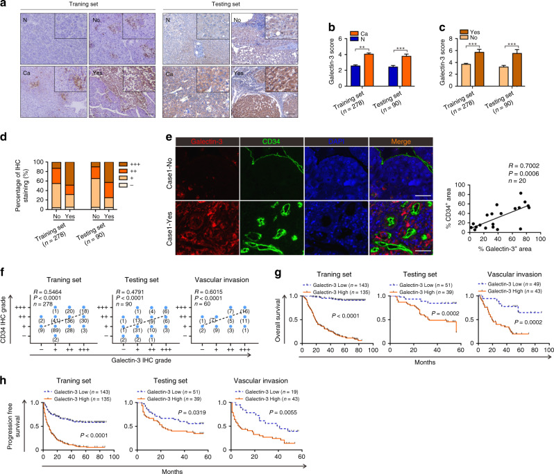
Background: High probability of metastasis limited the long-term survival of patients with hepatocellular carcinoma (HCC). Our previous study revealed that Galectin-3 was closely associated with poor prognosis in HCC patients.

Methods: The effects of Galectin-3 on tumour metastasis were investigated in vitro and in vivo, and the underlying biological and molecular mechanisms involved in this process were evaluated.

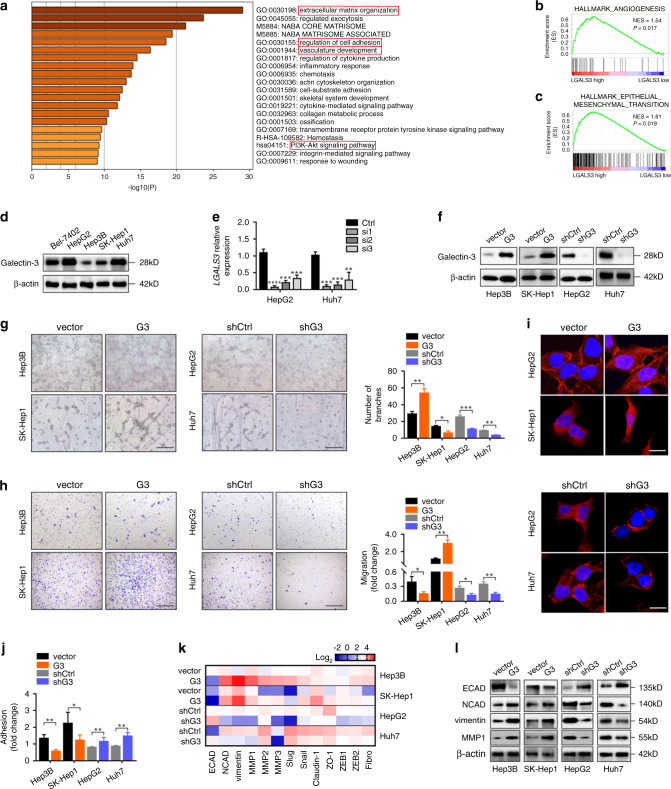
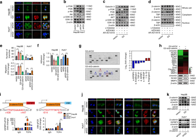
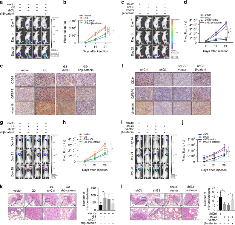
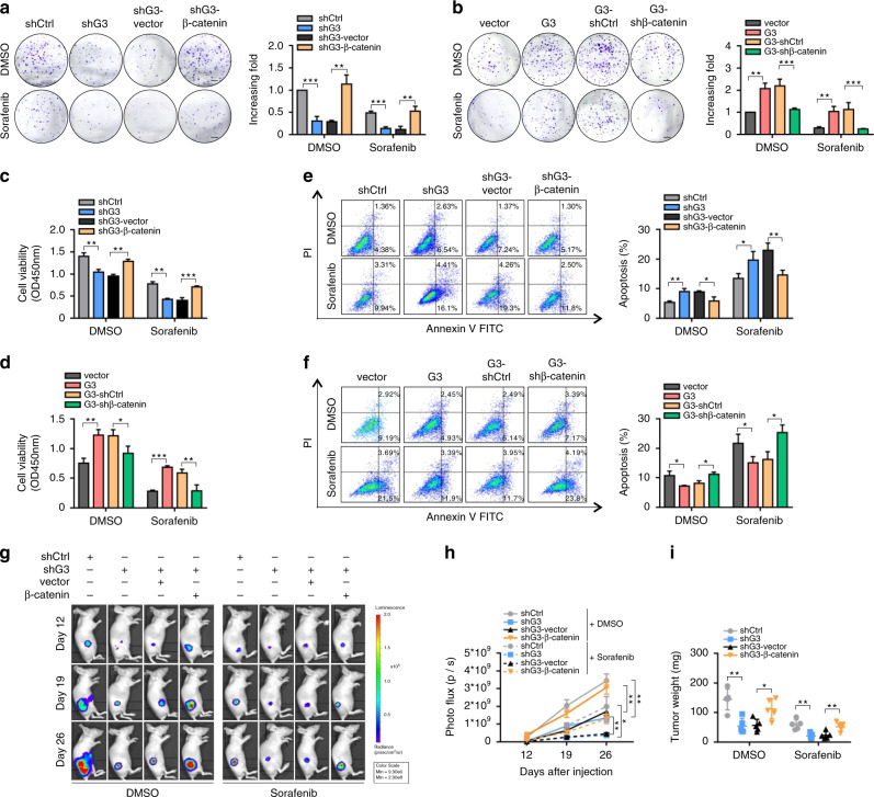
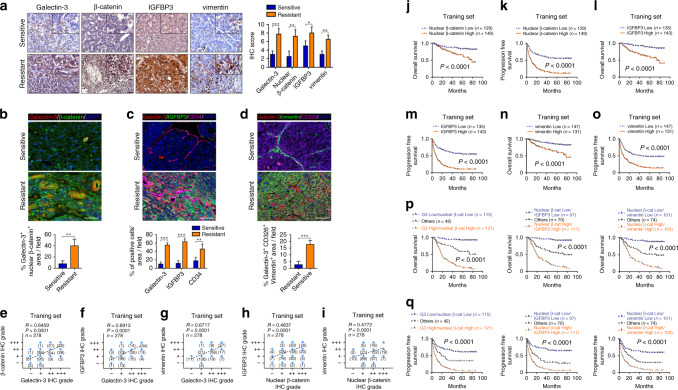
Results: Galectin-3 showed a close correlation with vascular invasion and poor survival in a large-scale study in HCC patients from multiple sets. Galectin-3 was significantly involved in diverse metastasis-related processes in HCC cells, such as angiogenesis and epithelial-to-mesenchymal transition (EMT). Mechanistically, Galectin-3 activated the PI3K-Akt-GSK-3β-β-catenin signalling cascade; the β-catenin/TCF4 transcriptional complex directly targeted IGFBP3 and vimentin to regulate angiogenesis and EMT, respectively. In animal models, Galectin-3 enhanced the tumorigenesis and metastasis of HCC cells via β-catenin signalling. Moreover, molecular deletion of Galectin-3-β-catenin signalling synergistically improved the antitumour effect of sorafenib.

Conclusions: The Galectin-3-β-catenin-IGFBP3/vimentin signalling cascade was determined as a central mechanism controlling HCC metastasis, providing possible biomarkers for predicating vascular metastasis and sorafenib resistance, as well as potential therapeutic targets for the treatment of HCC patients.


Conflict of interest statement

The authors declare no competing interests.

Figures

Fig. 1
Fig. 1. A high level of Galectin-3 expression is significantly associated with vascular invasion and poor survival in HCC patients.
ac IHC staining shows the expression level of Galectin-3 in HCC tissues and paired adjacent normal tissues, as well as in HCC tissues with or without vascular invasion in the training and testing sets. Scale bars, 100 μm. d The percentage of Galectin-3 IHC staining grade in patients with or without vascular invasion in the training and testing sets. e Tissue IF staining shows the co-expression pattern of Galectin-3 (red) and CD34 (green) in the HCC tumour microenvironment. DAPI, blue. Figure panel pairs representative images taken with different zooming options; Scale bars, 100 μm. f Spearman’s correlation analysis of the association between Galectin-3 and CD34 in the training (left), testing (middle), and vascular invasion (right) sets. g, h Survival analyses between “Galectin-3 high” and “Galectin-3 low” groups for the overall survival (g) and progression-free survival (h) based on IHC staining in the training (left), testing (middle), and vascular invasion (right) sets. N normal, Ca cancer, No without vascular invasion, Yes with vascular invasion. The results represent three independent experiments. **P < 0.01; ***P < 0.001.
Fig. 2
Fig. 2. Galectin-3 promotes the angiogenesis and EMT of HCC cells in vitro.
a GO analysis was performed according to the HCC mRNA sequencing data from TCGA to identify Galectin-3-associated GO terms; the top 20 terms are shown. b, c GSEA based on the HCC mRNA sequencing data from TCGA as performed to analyse the correlation between Galectin-3 and angiogenesis (b) and EMT (c). d Western blotting shows the expression of Galectin-3 in HCC cell lines, including Bel-7402, HepG2, Hep3B, SK-Hep1 and Hun7. e qPCR shows the mRNA expression of LGALS3 in Hep3B and SK-Hep1 cells transfected with siControl, siLGALS3#1, siLGALS3#2 or siLGALS3#3. f Western blotting shows the expression of Galectin-3 in Hep3B-vector, Hep3B-Galectin-3, SK-Hep1-vector, SK-Hep1-Galectin-3, HepG2-shControl, HepG2-shGalectin-3, Huh7-shControl and Huh7-shGalectin-3 cells. gj Tube formation assay (g), Transwell migration assay (h), IF assay for phalloidin (i), and cell adhesion assay (j) in Hep3B-vector, Hep3B-Galectin-3, SK-Hep1-vector, SK-Hep1-Galectin-3, HepG2-shControl, HepG2-shGalectin-3, Huh7-shControl and Huh7-shGalectin-3 cells. Scale bars in g, h, and i are 200 μm, 200 μm, and 20 μm, respectively. k PCR array shows the expression of EMT-related genes in Hep3B-vector, Hep3B-Galectin-3, SK-Hep1-vector, SK-Hep1-Galectin-3, HepG2-shControl, HepG2-shGalectin-3, Huh7-shControl and Huh7-shGalectin-3 cells. l Western blotting shows the expression of ECAD, NCAD, vimentin and MMP1 in Hep3B-vector, Hep3B-Galectin-3, SK-Hep1-vector, SK-Hep1-Galectin-3, HepG2-shControl, HepG2-shGalectin-3, Huh7-shControl and Huh7-shGalectin-3 cells. Ctrl control, G3 Galectin-3. The results represent three independent experiments. *P < 0.05; **P < 0.01; ***P < 0.001.
Fig. 3
Fig. 3. Activation of the PI3K-Akt-GSK-3β-β-catenin signalling cascade is essential for Galectin-3-induced angiogenesis and EMT of HCC cells.
a IF staining shows the colocalisation of β-catenin (green) and Galectin-3 (red) in Hep3B-vector, Hep3B-Galectin-3, Huh7-shControl and Huh7-shGalectin-3 cells. Scale bars, 20 μm. b Western blotting shows the expression of phospho-PI3K, PI3K, phospho-Akt, Akt, phospho-GSK-3β and GSK-3β in Hep3B-vector, Hep3B-Galectin-3, Huh7-shControl and Huh7-shGalectin-3 cells. c, d Hep3B-Galectin-3 cells were treated with DMSO, PF294002 (20 μmol/L), AZD5363 (10 μmol/L) or AR-A014418 (20 μmol/L). Western blotting shows the expression of Galectin-3, phospho-PI3K, PI3K, phospho-Akt, Akt, phospho-GSK-3β and GSK-3β (c). Western blotting of the expression of β-catenin in the whole cell, cytoplasm and nucleus (d). e, f Statistical diagrams of the tube formation assay, Transwell migration assay and cell adhesion assay. g, h Proteome angiogenesis array shows angiogenesis-related soluble proteins (g), and PCR array and western blotting show EMT-related genes/proteins (h) in Hep3B-shControl and Hep3B-shβ-catenin cells based on Galectin-3 overexpression. i Analysis of the IGFBP3 and VIM promoter identified a TCF4-binding site. Moreover, CHIP was performed using IgG and TCF4 antibodies, followed by qPCR in Hep3B-vector, Hep3B-Galectin-3, Huh7-shControl and Huh7-shGalectin-3 cells. j IF staining shows the colocalisation of β-catenin (green) and IGFBP3 (red) (left) or vimentin (red) (right) in Hep3B-vector, Hep3B-Galectin-3, Huh7-shControl and Huh7-shGalectin-3 cells. Scale bars, 20 μm. k Western blotting shows the expression of β-catenin, IGFBP3 and vimentin in Hep3B-vector, Hep3B-Galectin-3, Hep3B-Galectin-3-shControl and Hep3B-Galectin-3-shβ-catenin cells (upper) and Huh7-shControl, Huh7-shGalectin-3, Huh7-shGalectin-3-vector and Huh7-shGalectin-3-β-catenin cells (bottom). Ctrl control, G3 Galectin-3. The results represent three independent experiments. *P < 0.05; **P < 0.01; ***P < 0.001.
Fig. 4
Fig. 4. Galectin-3 facilitates tumorigenesis and lung metastasis of HCC cells in vivo via β-catenin signalling.
a, b Representative images and photo flux of tumour growth of Hep3B-vector, Hep3B-Galectin-3, Hep3B-Galectin-3-shControl and Hep3B-Galectin-3-shβ-catenin cells in BALB/c nude mice at day 7, 14 and 21. c, d Representative images and photo flux of tumour growth of Huh7-shControl, Huh7-shGalectin-3, Huh7-shGalectin-3-vector and Huh7-shGalectin-3-β-catenin cells in BALB/c nude mice at day 7, 14 and 21. e, f IHC staining shows the expression of CD34, IGFBP3 and vimentin in xenografts. Scale bars, 100 μm. g, h Representative images and photo flux of lung metastasis of Hep3B-vector, Hep3B-Galectin-3, Hep3B-Galectin-3-shControl and Hep3B-Galectin-3-shβ-catenin cells in BALB/c nude mice at day 14, 21 and 28. i, j Representative images and photo flux of lung metastasis of Huh7-shControl, Huh7-shGalectin-3, Huh7-shGalectin-3-vector and Huh7-shGalectin3-β-catenin cells in BALB/c nude mice at day 14, 21 and 28. k, l H&E staining shows micro-metastatic pulmonary nodules of Hep3B-vector, Hep3B-Galectin-3, Hep3B-Galectin-3-shControl, Hep3B-Galectin-3-shβ-catenin, Huh7-shControl, Huh7-shGalectin-3, Huh7-shGalectin-3-vector and Huh7-shGalectin-3-β-catenin cells in BALB/c nude mice. Scale bars, 100 μm. Ctrl control, G3 Galectin-3. The results represent three independent experiments. *P < 0.05; **P < 0.01; ***P < 0.001.
Fig. 5
Fig. 5. Molecular deletion of Galectin-3-β-catenin signalling synergistically improves the antitumour effect of sorafenib in vitro and in vivo.
af After cell adhesion, DMSO or sorafenib (20 μmol/mL) were administered every two days for the colony formation assay, (a, b), CCK8 assay (c, d) and apoptosis assay (e, f) in Hep3B-vector, Hep3B-Galectin-3, Hep3B-Galectin-3-shControl, Hep3B-Galectin-3-shβ-catenin, Huh7-shControl, Huh7-shGalectin-3, Huh7-shGalectin-3-vector and Huh7-shGalectin-3-β-catenin cells. Scale bars in a and b, 5 mm. g, h Representative images and photo flux of tumour growth of Hep3B-vector, Hep3B-Galectin-3, Hep3B-Galectin-3-shControl, Hep3B-Galectin-3-shβ-catenin, Huh7-shControl, Huh7-shGalectin-3 Huh7-shGalectin-3-vector and Huh7-shGalectin-3-β-catenin cells in BALB/c nude mice treated with DMSO or sorafenib at day 12, 19, and 26. i Mice were sacrificed at day 27. The tumour weights were measured. Ctrl control, G3 Galectin-3. The results represent three independent experiments. *P < 0.05; **P < 0.01; ***P < 0.001.
Fig. 6
Fig. 6. High-level Galectin-3-β-catenin-IGFBP3/vimentin axis is correlated with vascular invasion-mediated sorafenib resistance and poor prognosis in large-scale clinical samples.
ad Sorafenib-sensitive patients without vascular invasion and sorafenib-resistant patients with vascular invasion were selected from the training set. a IHC staining shows the expression levels of Galectin-3, β-catenin, IGFBP3 and vimentin. Scale bars, 50 μm. bd Tissue IF staining shows the co-expression pattern of Galectin-3 (red) and β-catenin (green) (b); Galectin-3 (red), IGFBP3 (green) and CD34 (magenta) (c); and Galectin-3 (red), vimentin (green) and CD326 (magenta) (d). DAPI, blue. Figure panel pairs the representative images taken with different zooming options; Scale bars, 100 μm. eh Spearman’s correlation analysis for the relationship between Galectin-3 and nuclear β-catenin (e); Galectin-3 and IGFBP3 (f); Galectin-3 and vimentin (g); nuclear β-catenin and IGFBP3 (h) and nuclear β-catenin and vimentin (i) in the training set according to IHC staining. jo Analysis of the differences between the high and low expression of nuclear β-catenin (j, k), IGFBP3 (l, m) and vimentin (n, o) for the OS and PFS based on IHC staining in the training set. p, q Kaplan–Meier analysis of the OS (p) and PFS (q) in the training set containing 278 HCC patients according to both Galectin-3 and nuclear β-catenin expression (left), nuclear β-catenin and IGFBP3 expression (middle) and nuclear β-catenin and vimentin expression (right), as assessed by IHC staining. No without vascular invasion, Yes with vascular invasion. β-cat β-catenin, G3 Galectin-3. The results represent three independent experiments. *P < 0.05; **P < 0.01; ***P < 0.001.

Similar articles

Cited by

References

    1. Torre LA, Bray F, Siegel RL, Ferlay J, Lortet-Tieulent J, Jemal A. Global cancer statistics, 2012. CA Cancer J. Clin. 2015;65:87–108. - PubMed
    1. Vogel A, Cervantes A, Chau I, Daniele B, Llovet JM, Meyer T, et al. Hepatocellular carcinoma: ESMO Clinical Practice Guidelines for diagnosis, treatment and follow-up. Ann. Oncol. 2019;30:871–873. - PubMed
    1. Morse MA, Sun W, Kim R, He AR, Abada PB, Mynderse M, et al. The role of angiogenesis in hepatocellular carcinoma. Clin. Cancer Res. 2019;25:912–920. - PubMed
    1. Siveen KS, Prabhu K, Krishnankutty R, Kuttikrishnan S, Tsakou M, Alali FQ, et al. Vascular endothelial growth factor (VEGF) signaling in tumour vascularization: potential and challenges. Curr. Vasc. Pharmacol. 2017;15:339–351. - PubMed
    1. Llovet JM, Di Bisceglie AM, Bruix J, Kramer BS, Lencioni R, Zhu AX, et al. Design and endpoints of clinical trials in hepatocellular carcinoma. J. Natl Cancer Inst. 2008;100:698–711. - PubMed

Publication types

MeSH terms