

The M-CSF receptor in osteoclasts and beyond
- PMID: 32801364
- PMCID: PMC8080670
- DOI: 10.1038/s12276-020-0484-z
The M-CSF receptor in osteoclasts and beyond
Abstract
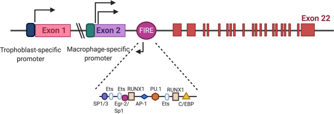
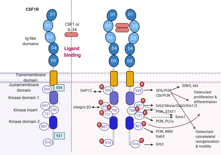
Colony-stimulating factor 1 receptor (CSF1R, also known as c-FMS) is a receptor tyrosine kinase. Macrophage colony-stimulating factor (M-CSF) and IL-34 are ligands of CSF1R. CSF1R-mediated signaling is crucial for the survival, function, proliferation, and differentiation of myeloid lineage cells, including osteoclasts, monocytes/macrophages, microglia, Langerhans cells in the skin, and Paneth cells in the intestine. CSF1R also plays an important role in oocytes and trophoblastic cells in the female reproductive tract and in the maintenance and maturation of neural progenitor cells. Given that CSF1R is expressed in a wide range of myeloid cells, altered CSF1R signaling is implicated in inflammatory, neoplastic, and neurodegenerative diseases. Inhibiting CSF1R signaling through an inhibitory anti-CSF1R antibody or small molecule inhibitors that target the kinase activity of CSF1R has thus been a promising therapeutic strategy for those diseases. In this review, we cover the recent progress in our understanding of the various roles of CSF1R in osteoclasts and other myeloid cells, highlighting the therapeutic applications of CSF1R inhibitors in disease conditions.
Conflict of interest statement
The authors declare that they have no conflict of interest.
Figures
Similar articles
-
CSF-1 receptor signaling in myeloid cells.Cold Spring Harb Perspect Biol. 2014 Jun 2;6(6):a021857. doi: 10.1101/cshperspect.a021857. Cold Spring Harb Perspect Biol. 2014. PMID: 24890514 Free PMC article. Review.
-
Protein kinase A inhibition of macrophage maturation is accompanied by an increase in DNA methylation of the colony-stimulating factor 1 receptor gene.Immunology. 2016 Oct;149(2):225-37. doi: 10.1111/imm.12641. Epub 2016 Aug 16. Immunology. 2016. PMID: 27353657 Free PMC article.
-
Small-molecule CSF1R kinase inhibitors; review of patents 2015-present.Expert Opin Ther Pat. 2021 Feb;31(2):107-117. doi: 10.1080/13543776.2021.1839414. Epub 2020 Oct 28. Expert Opin Ther Pat. 2021. PMID: 33108917 Review.
-
Regulation of Embryonic and Postnatal Development by the CSF-1 Receptor.Curr Top Dev Biol. 2017;123:229-275. doi: 10.1016/bs.ctdb.2016.10.004. Epub 2016 Dec 1. Curr Top Dev Biol. 2017. PMID: 28236968 Free PMC article. Review.
-
Therapeutic applications of macrophage colony-stimulating factor-1 (CSF-1) and antagonists of CSF-1 receptor (CSF-1R) signaling.Blood. 2012 Feb 23;119(8):1810-20. doi: 10.1182/blood-2011-09-379214. Epub 2011 Dec 20. Blood. 2012. PMID: 22186992 Review.
Cited by
-
Rare coding variants in NOX4 link high ROS levels to psoriatic arthritis mutilans.EMBO Mol Med. 2024 Mar;16(3):596-615. doi: 10.1038/s44321-024-00035-z. Epub 2024 Feb 20. EMBO Mol Med. 2024. PMID: 38379095 Free PMC article.
-
Development of an optogenetics tool, Opto-RANK, for control of osteoclast differentiation using blue light.Sci Rep. 2024 Jan 19;14(1):1749. doi: 10.1038/s41598-024-52056-w. Sci Rep. 2024. PMID: 38242937 Free PMC article.
-
RANKL-dependent osteoclast differentiation and gene expression in bone marrow-derived cells from adult mice is sexually dimorphic.Bone Rep. 2023 Jul 1;19:101697. doi: 10.1016/j.bonr.2023.101697. eCollection 2023 Dec. Bone Rep. 2023. PMID: 37485233 Free PMC article.
-
Leveraging mice with diverse microbial exposures for advances in osteoimmunology.Front Endocrinol (Lausanne). 2023 May 10;14:1168552. doi: 10.3389/fendo.2023.1168552. eCollection 2023. Front Endocrinol (Lausanne). 2023. PMID: 37251680 Free PMC article.
-
Live Cell Sorting of Differentiated Primary Human Osteoclasts Allows Generation of Transcriptomic Signature Matrix.Res Sq [Preprint]. 2025 Apr 1:rs.3.rs-6157400. doi: 10.21203/rs.3.rs-6157400/v1. Res Sq. 2025. PMID: 40235499 Free PMC article. Preprint.
References
-
- Tsukasaki M, Takayanagi H. Osteoimmunology: evolving concepts in bone-immune interactions in health and disease. Nat. Rev. Immunol. 2019;19:626–642. - PubMed
-
- Boyle WJ, Simonet WS, Lacey DL. Osteoclast differentiation and activation. Nature. 2003;423:337–342. - PubMed
-
- Novack DV, Teitelbaum SL. The osteoclast: friend or foe? Annu. Rev. Pathol. 2008;3:457–484. - PubMed
Publication types
MeSH terms
Substances
Grants and funding
LinkOut - more resources
Full Text Sources
Other Literature Sources
Research Materials
Miscellaneous
