

Intracardiac Echocardiography as a Guide for Transcatheter Closure of Patent Ductus Arteriosus
- PMID: 32802008
- PMCID: PMC7414339
- DOI: 10.1155/2020/5147193
Intracardiac Echocardiography as a Guide for Transcatheter Closure of Patent Ductus Arteriosus
Abstract
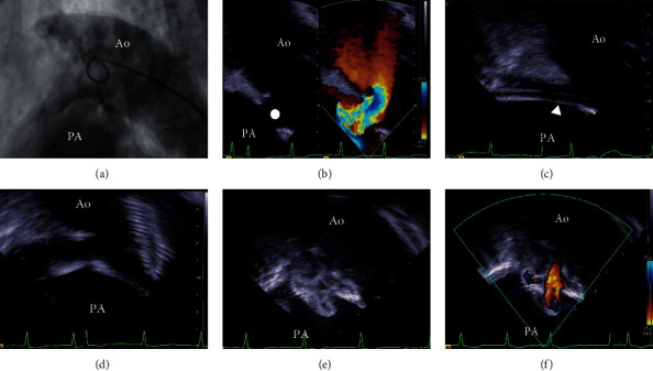
Background: Transcatheter closure of patent ductus arteriosus (TC-PDA), conventionally guided by aortography, has become the standard treatment of this disease. The purposes of this study were to evaluate whether intracardiac echocardiography (ICE) may be used for measuring PDA size and be used as a guide for TC-PDA.
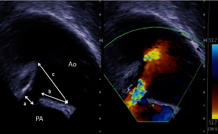
Methods: This study had 2 phases. In phase 1, we compared the measurements of PDA size: pulmonary artery side diameter (PA-D), length, and aortic side diameter (Ao-D) of PDA, as measured by ICE with those measured by aortography or cardiac computed tomography (AoG/CCT) in 23 patients who underwent TC-PDA. In phase 2, we compared the demographics, fluoroscopic time, contrast volume, and complications of the TC-PDAs between 10 adult patients with ICE guidance and 16 without it.
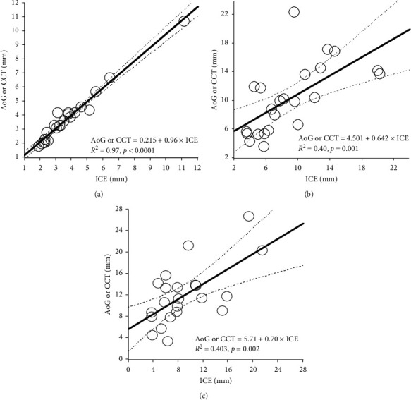
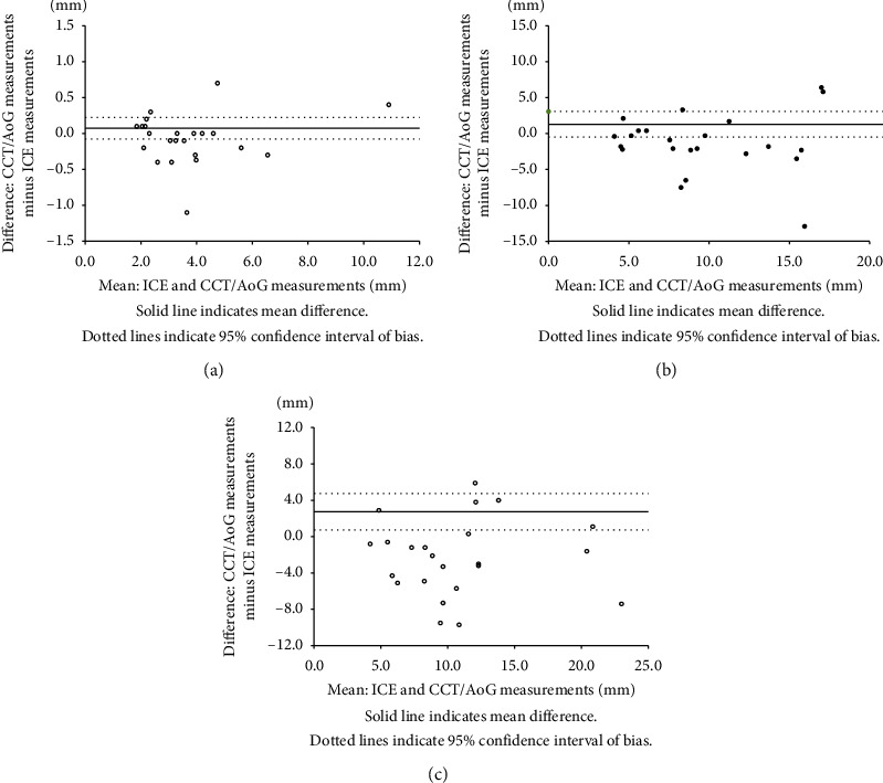
Results: In phase 1, we found great correlation and agreement between ICE and AoG/CCT in PA-D (r = 0.985, bias -0.077 to 0.224), but moderate to poor correlation and agreement in length (r = 0.653, bias -0.491 to 3.065) and Ao-D (r = 0.704, bias 0.738 to 4.732), respectively. Nevertheless, all patients underwent successful TC-PDA with ICE guidance that allowed us to continuously monitor the whole process. In phase 2, TC-PDA required a significantly lower contrast volume with ICE guidance than without it, and there was no significant difference in the remaining variables between the 2 groups.
Conclusion: ICE is comparable to AoG/CCT in providing accurate PA-D of the PDA and may be a safe alternative to guide TC-PDA as compared to conventional aortography.
Copyright © 2020 Hironaga Yoshimoto et al.
Conflict of interest statement
The authors declare that they have no conflicts of interest with the contents of this article.
Figures

Similar articles
-
Trans-pulmonary echocardiography as a guide for device closure of patent ductus arteriosus.Catheter Cardiovasc Interv. 2015 Aug;86(2):264-70. doi: 10.1002/ccd.25879. Epub 2015 Feb 25. Catheter Cardiovasc Interv. 2015. PMID: 25676054
-
Echocardiographic versus Angiographic Measurement of the Patent Ductus Arteriosus in Extremely Low Birth Weight Infants and the Utility of Echo Guidance for Transcatheter Closure.J Am Soc Echocardiogr. 2021 Oct;34(10):1086-1094. doi: 10.1016/j.echo.2021.06.005. Epub 2021 Jun 15. J Am Soc Echocardiogr. 2021. PMID: 34139301
-
Transthoracic echocardiography as an alternative major guidance to angiography during transcatheter closure of patent ductus arteriosus: technical feasibility and clinical relevance.Pediatr Cardiol. 2015 Jan;36(1):14-9. doi: 10.1007/s00246-014-0956-9. Epub 2014 Jul 29. Pediatr Cardiol. 2015. PMID: 25070385
-
Morphologic characterization of the patent ductus arteriosus in the premature infant and the choice of transcatheter occlusion device.Catheter Cardiovasc Interv. 2016 Feb 1;87(2):310-7. doi: 10.1002/ccd.26287. Epub 2015 Nov 3. Catheter Cardiovasc Interv. 2016. PMID: 26525611
-
Closure of the patent ductus arteriosus with the new duct occluder II additional sizes device.Catheter Cardiovasc Interv. 2012 Jun 1;79(7):1169-74. doi: 10.1002/ccd.23477. Epub 2012 Mar 15. Catheter Cardiovasc Interv. 2012. PMID: 22422478
Cited by
-
Intracardiac echocardiography Chinese expert consensus.Front Cardiovasc Med. 2022 Oct 6;9:1012731. doi: 10.3389/fcvm.2022.1012731. eCollection 2022. Front Cardiovasc Med. 2022. PMID: 36277762 Free PMC article. Review.
-
Evaluation of Cardiac Function Characteristics after Patent Ductus Arteriosus Closure in Children and Adults by Echocardiographic Data.Comput Math Methods Med. 2022 Jan 28;2022:1310841. doi: 10.1155/2022/1310841. eCollection 2022. Comput Math Methods Med. 2022. Retraction in: Comput Math Methods Med. 2023 Nov 1;2023:9768428. doi: 10.1155/2023/9768428. PMID: 35126616 Free PMC article. Retracted.
References
-
- Porstmann W., Wierny L., Warnke H. Closure of persistent ductus arteriosus without thoracotomy. German Medical Monthly. 1967;12(6):259–261. - PubMed
-
- Baumgartner H., Bonhoeffer P., De Groot N. M., et al. ESC Guidelines for the management of grown-up congenital heart disease (new version 2010) European Heart Journal. 2010;31(23):2915–2957. - PubMed
MeSH terms
LinkOut - more resources
Full Text Sources
