Review
. 2020 Aug 7;30(32):2000639.
doi: 10.1002/adfm.202000639. Epub 2020 Jun 18.

Spatiotemporally Controlled Photoresponsive Hydrogels: Design and Predictive Modeling from Processing through Application

Affiliations
Review

Spatiotemporally Controlled Photoresponsive Hydrogels: Design and Predictive Modeling from Processing through Application

Hongyuan Zhu et al. Adv Funct Mater. .

Abstract

Photoresponsive hydrogels (PRHs) are soft materials whose mechanical and chemical properties can be tuned spatially and temporally with relative ease. Both photo-crosslinkable and photodegradable hydrogels find utility in a range of biomedical applications that require tissue-like properties or programmable responses. Progress in engineering with PRHs is facilitated by the development of theoretical tools that enable optimization of their photochemistry, polymer matrices, nanofillers, and architecture. This review brings together models and design principles that enable key applications of PRHs in tissue engineering, drug delivery, and soft robotics, and highlights ongoing challenges in both modeling and application.

Keywords: hydrogels; mechanical properties; models; photodegradation; photo‐crosslinking.


Conflict of interest statement

The authors declare no conflict of interest.

Figures
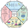
Figure 1
Figure 1
Basic design principles and promising applications of PRHs with tuned mechanical properties.
Figure 2
Figure 2
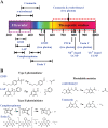
A) Absorption wavelength distribution and B) reaction schemes of widely used biocompatible photosensitive compounds. When photoresponsive hydrogels are irradiated with light, photoinitiators can trigger photo‐crosslinking, while photolabile moieties can trigger photodegradation. The absorption wavelengths of these photosensitive compounds are located mainly in the ultraviolet and visible regions. To achieve light control in biologically benign regions (600–1000 nm), NIR excitation techniques based on two‐photon absorption and upconversion nanoparticles have been developed.
Figure 3
Figure 3
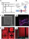
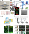
Two‐photon absorption (TPA) techniques for NIR‐engineering PRHs. A) Schematic energy‐level diagram of TPA. A molecule at ground state (g) could be excited to excited state (f) by simultaneously absorbing two photons with energy E 1, E 2, (E 1 could equal to E 2). After excitation, the molecule relaxes to lowest vibronic level (r), and then returns to the ground state by radiative or irradiative pathways. B) Typical setup for two‐photon PRH fabrication. Reproduced with permission.[ 47 ] Copyright 2018, Royal Society of Chemistry. C) Woodpile scaffold fabricated by two‐photon photopolymerization. Reproduced with permission.[ 51a ] Copyright 2017, Elsevier. a) Scanning‐electron microscopy images of a scaffold. b) Fluorescence image of the scaffold and seeded cells. D) 3D channels for cell culture fabricated by two‐photon degradation. Reproduced with permission.[ 25a ] Copyright 2011, Nature Publishing Group. a) Confocal images of a 3D structure (scale bar: 100 µm). b) Fluorescence image of a channel with seeded cells (scale bar: 100 µm).
Figure 4
Figure 4
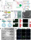
Upconversion nanoparticle (UCNP)‐assisted NIR‐engineering of PRHs. A) Schematic energy‐level diagram of an Yb3+/Er3+ ion pair. Reproduced with permission.[ 59 ] Copyright 2015, Royal Society of Chemistry. B) Schematic of UCNP‐assisted NIR‐induced photochemistry. Reproduced with permission.[ 55 ] Copyright 2017, Royal Society of Chemistry. C) UCNP‐assisted 3D fabrication. Reproduced under the terms of the CC‐BY License.[ 54a ] Copyright 2018, the Authors. Published by Nature Publishing Group. a) Schematic of the experimental setup. b) Luminescent voxel formation in resin‐contained UCNPs under CW NIR light illumination. c) Scanning electron microscopy image of a 3D polymer microstructure obtained by UCNP‐assisted 3D fabrication. D) NIR‐controlled cell adhesion using UCNPs. Reproduced with permission.[ 66a ] Copyright 2014, American Chemical Society. a) Schematic illustration. b,c) Fluorescence images of NIR light‐induced cells released on substrate with b) and without c) UCNPs (scale bar: 100 µm). E. NIR‐controlled differentiation of MSCs using UCNPs. Reproduced with permission.[ 67 ] Copyright 2018, Wiley‐VCH. a) The matrix was modified by two distinct polymers: P1, which is (ONB‐PEG), and P2, which is (RGD‐PEG). P1 is photocleavable and can block interaction between cells and the substrate, while P2 can anchor the cells. NIR irradiation could trigger UCNP‐assisted detachment of P1, and change cell–matrix interactions. b) Immunofluorescence imaging of markers for osteogenic (RUNX2, green) differentiation under matrix with various NIR irradiation doses (scale bar: 50 µm).
Figure 5
Figure 5
Polymer concentration‐dependent Young's modulus plot for PRHs with a range of polymer matrices. Data are from reports on gelatin‐derived,[ 23 , 80 , 81 ] hyaluronic acid (HA)‐derived,[ 85 , 86 , 87 , 88 ] polyacrylamide (PAAm)‐derived,[ 94 ] poly ethylene glycol (PEG)‐derived,[ 25 , 97 , 98 ] and poly vinyl alcohol (PVA)‐derived [ 51b,c ] PRHs. The measured shear modulus (G) in these references is transformed into Young's modulus (E) by E = 2(1+ν)G, where ν is Poisson's ratio, which was assumed to be 0.5.
Figure 6
Figure 6
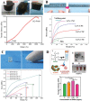
Reinforcement of PRHs by nanoparticle fillers. A) Photographs and tensile stress–strain curves of a graphene oxide (GO)‐doped polyacrylamide (PAAm) hydrogel, where Ca2+ is used as the crosslinker of GO sheets. The composite hydrogel is much more ductile and stiff than an undoped PAAm hydrogel. Reproduced with permission.[ 128 ] Copyright 2013, Wiley‐VCH. B) Photographs and tensile stress–strain curves of a cellulose nanocrystal (CNC)‐doped PAAm hydrogel. With increasing CNC nanofiller content, the composite hydrogels increased in Young's modulus and fracture strength. Reproduced with permission.[ 136 ] Copyright 2013, American Chemical Society. C) Photographs and compressive stress–strain curves of a clay nanoparticle‐doped poly N‐acryloyl glycinamide (PNAGA) hydrogel. The clay nanofiller increased the compressive modulus and strength of the hydrogel. Reproduced with permission.[ 149 ] Copyright 2017, American Chemical Society. D) Preparation of GelMA‐coated gold nanorods (G‐GNRs)/GelMA hydrogels. TEM image and photographs of G‐GNRs in solution, and Young's moduli of G‐GNRs/GelMA hydrogels at various concentrations of G‐GNRs. Reproduced with permission.[ 108e ] Copyright 2017, Wiley‐VCH.
Figure 7
Figure 7
3D printing methods for PRHs. A) Representative light‐based printing techniques. a) Fabrication of patterned hydrogel structures by photomask‐based stereolithography (scale bar: 2 mm). Reproduced with permission.[ 181 ] Copyright 2013, Wiley‐VCH. b) Fabrication of a multilayer cell‐laden hydrogel by laser‐based stereolithography (scale bar: 1 mm). Reproduced with permission.[ 182a ] Copyright 2010, Royal Society of Chemistry. c) Fabrication via digital micromirror device (DMD)‐projection‐based stereolithography and SEM images of microstructured wells (scale bar: 2 mm). Reproduced with permission.[ 179 ] Copyright 2012, Wiley‐VCH. B) Representative ink‐based printing techniques. a) Inkjet printing of cell‐loaded photo‐crosslinkable hydrogels and the resulting even cell distribution within the hydrogels (scale bar: 100 µm). Reproduced with permission.[ 191c ] Copyright 2015, Springer. b) Extrusion printing of photo‐crosslinkable hydrogels by three different fabrication strategies (pre‐, post‐, in situ photo‐crosslinking) with fibers printed by an in situ photo‐crosslinking strategy (scale bar: 500 µm). Reproduced with permission.[ 197 ] Copyright 2017, Wiley‐VCH.
Figure 8
Figure 8
Basic principles for modeling the elasticity of photo‐crosslinkable hydrogels. There are three tandem relationships underlying the evolution of mechanics in photo‐crosslinkable hydrogels: the irradiation time‐dependent degree of conversion (DoC), DoC‐dependent network topology, and network topology‐dependent elasticity. Theoretical methods have been developed for each relationship. From left to right, adapted with permission. [ 206 ] Copyright 2018, Elsevier.[ 215a ] Copyright 2017, Wiley‐VCH.[ 199b ] Copyright 2016, American Association for the Advancement of Science.
Figure 9
Figure 9
Development of models for the elasticity of photodegradable hydrogels. In 2009, a simple exponential function was introduced to express the irradiation time‐dependent elastic moduli of photodegradable hydrogels. Adapted with permission.[ 32a ] Copyright 2009, American Association for the Advancement of Science. Optical attenuation and mass loss were incorporated into models for optically thick hydrogels in 2013. Adapted with permission.[ 238 ] Copyright 2013, Wiley‐VCH. Models accounting for degradation byproduct diffusion were developed in 2017. Adapted with permission.[ 239 ] Copyright 2017, Wiley‐VCH.
Figure 10
Figure 10
Relationships between the ratio of Young's modulus of nanocomposite (Ec) and matrix (Em) and volume fraction of fillers (φ) for different nanocomposites. A) Carbon nanotube filled PRHs.[ 108 , 129 ] B) Cellulose reinforced PRHs.[ 108 , 138 , 139 ] C) Clay particulate reinforced PRHs.[ 144 , 145 , 148 ] D) Metal particle reinforced PRHs.[ 108 , 152 ] The dots are representative experimental data obtained from the literature. The estimates and bounds shown are two‐phase homogenization results for a PRH matrix (Em = 1 kPa, νm = 0.49) reinforced by rigid spherical inclusions (Ef = 1 TPa, νf = 0.3), where νm and are Poisson's ratios of matrix and filler, respectively. In many cases, predicted stiffening rises above thermodynamic limits for conventional composites due to surface energy effects such as recrystallization that arise from nanofillers.
Figure 11
Figure 11
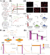
Cell culture platforms based on PRH with temporally controlled elasticity. A) A representative matrix‐stiffening strategy for PRHs used in cell culture. Reproduced under the terms of the CC‐BY License.[ 278 ] Copyright 2012, the Authors. Published by Nature Publishing Group. a) Schematic of the sequential crosslinking process in methacrylated hyaluronic acid (MeHA). b) Rheology profiles over the course of the hydrogel formation process. c) Atomic force microscopy estimates of the Young's moduli of MeHA substrates at various DTT concentrations (scale bar: 100 µm). d) Fluorescence images of MSCs cultured on MeHA substrates (soft, soft‐to‐stiff, stiff) for 1 and 2 days. e) Distributions of cell areas when cultured on the static and dynamic substrates in (d). B) Representative softening strategy for PRHs used in cell culture. Reproduced with permission.[ 283b ] Copyright 2014, Nature Publishing Group. a) Schematic of the softening of a photodegradable hydrogel. b) Young's moduli of photodegradable hydrogel at various light exposure doses. c) Cell responses (nuclear co‐localization of YAP and RUNX2) to mechanical dosing on stiff hydrogels.
Figure 12
Figure 12
Cell culture platforms based on PRHs with spatially heterogeneous elasticity. A) Representative cell migration study based on mechanically patterned substrates. Reproduced with permission.[ 290b ] Copyright 2013, Wiley‐VCH. a) Three different strengths of stiffness gradients (step, pathological, and physiological), and b) velocities of mesenchymal stem cells (MSCs) migrating along these gradients. c) Kernel density estimation of cell velocities on the three stiffness gradients. B) High‐throughput cell behavior study based on mechanically patterned substrates. Reproduced under the terms of the CC‐BY License.[ 169a ] Copyright 2015, the Authors. Published by Nature Publishing Group. a) Orthogonal gradients of both stiffness and fibronectin concentrations produced by lithography. b) Statistical results of osteogenic staining of MSCs on orthogonal gradient hydrogels. c) Statistical results of adipogenic staining of MSCs on the orthogonal gradient hydrogels. C) A representative study showing cellular responses to subcellular elastic heterogeneity arising on mechanically patterned substrates. Reproduced under the terms of the CC‐BY License.[ 174b ] Copyright 2016, the Authors. Published by National Academy of Sciences. a) MSCs on mechanically patterned hydrogel surfaces with various stiff‐to‐soft ratios. Black indicates stiff regions, and white indicates soft regions. b) Dependence of cell morphology (cellular circularity) on substrate composition for substrates with various percentages of stiff regions and a range of patterns.
Figure 13
Figure 13
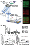
Hepatic triculture model constructed from multiple cell‐loaded PRHs. Reproduced with permission.[ 183 ] Copyright 2016, National Academy of Sciences. A) Schematic of fabrication processes a) and fluorescence images b) of a printed triculture hepatic model. In green are human‐induced pluripotent stem cell (hiPSC)‐derived hepatic progenitor cells (HPCs), and in red are supporting cells (scale bar: 500 µm). B) Gene expression a), albumin secretion levels b), and urea secretion levels c) of the hepatic model. Results showed more mature gene expression and better anabolic and catabolic functionality of hiPSCs–HPCs in the printed triculture models than in other culture conditions.
Figure 14
Figure 14
Implantable biomaterials based on PRHs. A) PRH‐based 3D‐printed hybrid scaffold for cartilage defect repair. Reproduced with permission.[ 299 ] Copyright 2018, Wiley‐VCH. a) Design and application of the hybrid scaffold. After a 3D printed support structure was placed in the defect, a cell‐laden photo‐crosslinkable hydrogel was injected into the support structure and polymerized via light. b) Representative histological images of stained sulfated glycosaminoglycans in empty and scaffold‐filled porcine chondral defects under either free swelling or dynamic strain loading after 4‐week culture. Here, * means the position of the defect or scaffold (scale bar: 100 µm). c) Semiquantification of the width of degenerated tissue in empty and scaffold‐filled defects under free swelling (solid) and dynamic strain loading (striped). These results indicate that the PRH‐based scaffold could prevent degeneration of cartilage adjacent to a defect. B) PRH‐based nucleus pulposus (NP) regeneration strategy. Reproduced with permission.[ 300b ] Copyright 2017, Elsevier. a) Schematic showing a nucleus pulposus cell (NPC)‐laden photo‐crosslinkable precursor of an interpenetrating network (IPN) hydrogel injected into the NP cavity and then crosslinked by a minimally invasive illumination device. b) Representative hematoxylin and eosin stained tissue of the untreated (control) and cell‐laden hydrogel‐treated (IPN Opt + NPC) porcine disc degeneration model at 12 weeks after implantation (scale bar: black is 2000 µm, blue is 100 µm). c) Histological score obtained from untreated (control), cell‐laden hydrogel (IPN Opt + NPC), hydrogel only (IPN Opt), and cell only (NPC + PBS) groups at 12 weeks after implantation. Results demonstrate that the photo‐crosslinkable IPN hydrogel facilitated regeneration of porcine degenerative NPs. C) PRH‐based soft tissue restoration strategy. Reproduced with permission.[ 39c ] Copyright 2011, American Association for the Advancement of Science. a) Schematic showing transdermal photo‐crosslinking of injected PEGDA–HA hydrogels. b) Magnetic resonance imaging (MRI) of implanted PEGDA–HA hydrogels in human abdominal skin at days 0 and 84. c) Persistent height of the implanted hydrogel with various compositions, showing effectiveness of this photo‐crosslinkable hydrogel in the restoration of soft tissue.
Figure 15
Figure 15
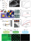
Drug delivery strategies based on PRHs. A) Prolonged drug release strategy based on abaloparatide‐loaded GelMA hydrogels. Reproduced with permission.[ 310 ] Copyright 2019, Wiley‐VCH. a) Schematic for in vivo treatment of bone defect by loaded GelMA hydrogels. b) SEM image of dried abaloparatide‐loaded GelMA hydrogels (scale bar: 10 µm). c) In vitro drug release profiles of the hydrogels. B) Methacrylated hyaluronic acid (MeHA)‐coated mesoporous silica nanoparticles (MSN) used for ultrasound treatment. Reproduced with permission.[ 315 ] Copyright 2017, Royal Society of Chemistry. a) Schematic of targeted delivery and synergic sonodynamic therapy and chemotherapy of tumor by doxorubicin‐loaded MSN–HA (DOX@MSN–HA). b) TEM images of MSN and DOX@MSN–HA (scale bar: 50 nm). c) Drug release profile of DOX from DOX@MSN and DOX@MSN–HA without hyaluronidase and sonication. C) 3D printed oral dosage form based on PEGDA hydrogel. Reproduced with permission.[ 317a ] Copyright 2016, Elsevier. a) Laser‐based stereolithography platform for manufacturing drug‐loaded tablets. b) Photograph and SEM image of 4‐aminosalicylic acid (4‐ASA)‐loaded PEGDA hydrogel. c) Drug release profile of 4‐ASA from printed tablets under dynamic pH conditions. D) Sequentially triggered release strategy based on multiple stimuli, namely light‐ and chemical‐induced cleavage in hydrogels (scale bar: 1 mm). Reproduced with permission.[ 320 ] Copyright 2019, Royal Society of Chemistry.
Figure 16
Figure 16
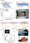
Electrical gauges based on PRHs. A) A NaCl containing ionic conductive PAAm hydrogel and its applications. Reproduced with permission.[ 324 ] Copyright 2013, American Association for the Advancement of Science. a) Construction and working principles of an electrical actuator formed by ionic hydrogels and dielectric elastomer. b) Expanded area of the electrical actuator under varying applied voltages on electrodes. c) A transparent loudspeaker generated from the electrical actuator. B) Gold nanowire (Au NW)‐filled conductive PAAm hydrogel and its applications. Reproduced with permission.[ 325c ] Copyright 2018, Wiley‐VCH. a) Working principles of a pressure sensor based on the conductive hydrogel. Pressure drives inner‐rib and between‐rib contact of Au NWs in the hydrogel sensor, reducing the resistance of the conductive hydrogels. b) Representative current responses and pressure sensitivities of the pressure sensors. c) Photographs of a wearable pressure sensor constructed from the conductive hydrogel.
Figure 17
Figure 17
Implantable sensor for marine organisms based on PRHs. Reproduced with permission. [ 330 ] Copyright 2019, American Chemical Society. A) Design schematic for implantable hydrogel sensors for physiological monitoring of marine organisms. B) Photograph of the fluorescent hydrogel sensor, which consists of DNA‐wrapped SWNTs and PEGDA (scale bar: 0.5 mm). C) In vitro tests of the sensor showing that the fluorescent signal of the hydrogel sensor decreases with step increases in riboflavin concentration. D) Overlay of brightfield and fluorescence images of a fluorescent hydrogel implanted beneath the skin of Galeus melastomus (scale bar: 20 mm).
Figure 18
Figure 18
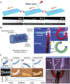
Actuators based on PRHs. A) Humidity‐driven actuator based on photo‐crosslinked PEGDA hydrogel films. Reproduced with permission.[ 333 ] Copyright 2017, Wiley‐VCH. When water vapor is applied to the film, the upper side absorbs the water and swells, which induces bending of the film. When water evaporates, the film is gradually stretched, which leads the device to move forward. B) Hydraulic hydrogel actuator fabricated from photo‐crosslinkable PAAm hydrogels. Reproduced under the terms of the CC‐BY License.[ 334 ] Copyright 2017, the Authors. Published by Nature Publishing Group. Inflation by water influx triggers the originally straight hydrogel to bend into a circle, and withdrawal of water restores its straight shape. C) Ion‐responsive composite hydrogel based on photo‐crosslinkable PEGDA and PAA hydrogels. Reproduced with permission.[ 335 ] Copyright 2019, Royal Society of Chemistry. Cations (Fe3+) trigger the crosslinking of anionic polymer chains (PAA), which results in contraction of the hydrogels. The mismatched contraction of the ion‐responsive layer (PEGDA) and non‐ion‐responsive layer (PEGDA–PAA) could result in bending or twisting actuation, which can further be used to design grippers (scale bar: 5 mm).
Figure 19
Figure 19
Robots based on PRHs. A) Soft electronic fish with PRH‐based components. Reproduced under the terms of the CC‐BY License.[ 321a ] Copyright 2017, the Authors. Published by American Association for the Advancement of Science. a) Design of the robot fish muscle, composed of an ionic hydrogel and dielectric elastomer. b) Tilted view showing the entire construction of the electronic fish. B) Soft microswimmers fabricated from PRHs (scale bar: 2 mm). Reproduced under the terms of the CC‐BY License.[ 337 ] Copyright 2016, the Authors. Published by Nature Publishing Group. a) The soft microswimmer was fabricated by photopatterning of magnetic hydrogel. b) A moving microswimmer driven by rotating uniform magnetic fields (scale bar: 5 mm). C) Bioinspired soft robot constructed from cell‐loaded PRHs. Reproduced with permission.[ 338 ] Copyright 2018, Wiley‐VCH. a) Construction scheme showing the layer‐by‐layer structure of the bioinspired soft robot. b) Photograph and SEM image showing the patterns of PEGDA and CNT/GelMA hydrogels on the robot. c) Photograph showing the cell‐actuating robots at over a contraction cycle.

Similar articles

Cited by

References

    1. Uto K., Tsui J. H., DeForest C. A., Kim D.‐H., Prog. Polym. Sci. 2017, 65, 53. - PMC - PubMed
    1. Wechsler M. E., Stephenson R. E., Murphy A. C., Oldenkamp H. F., Singh A., Peppas N. A., Biomed. Microdevices 2019, 21, 31. - PMC - PubMed
    1. a) Wallin T. J., Pikul J., Shepherd R. F., Nat. Rev. Mater. 2018, 3, 84;
    2. b) Truby R. L., Lewis J. A., Nature 2016, 540, 371; - PubMed
    3. c) Wang R., Sing M. K., Avery R. K., Souza B. S., Kim M., Olsen B. D., Acc. Chem. Res. 2016, 49, 2786; - PubMed
    4. d) Tomatsu I., Peng K., Kros A., Adv. Drug Delivery Rev. 2011, 63, 1257; - PubMed
    5. e) Ma Y., Lin M., Huang G., Li Y., Wang S., Bai G., Lu T. J., Xu F., Adv. Mater. 2018, 30, 1705911; - PubMed
    6. f) Dong Y., Jin G., Hong Y., Zhu H., Lu T. J., Xu F., Bai D., Lin M., ACS Appl. Mater. Interfaces 2018, 10, 12374. - PubMed
    1. Zhang Y. S., Khademhosseini A., Science 2017, 356, eaaf3627. - PMC - PubMed
    1. Matsuda T., Kawakami R., Namba R., Nakajima T., Gong J. P., Science 2019, 363, 504. - PubMed

LinkOut - more resources