. 2020 Jul 31:2020:5264205.
doi: 10.1155/2020/5264205. eCollection 2020.

Buyang Huanwu Decoction Promotes Angiogenesis after Cerebral Ischemia by Inhibiting the Nox4/ROS Pathway

Affiliations

Buyang Huanwu Decoction Promotes Angiogenesis after Cerebral Ischemia by Inhibiting the Nox4/ROS Pathway

Jian Shen et al. Evid Based Complement Alternat Med. .

Abstract

Background: Buyang Huanwu decoction (BYHWD), an important traditional Chinese medicine (TCM), has been used clinically for centuries for the treatment of various diseases. The study aims to explore the BYHWD effects on angiogenesis and neuroprotection after cerebral ischemia/reperfusion (CI/R) injury in rats and to explore the underlying angiogenic roles and mechanisms of BYHWD in hydrogen peroxide (H2O2) induced oxidative stress in human umbilical vein endothelial cells (HUVECs) model.

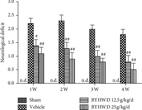
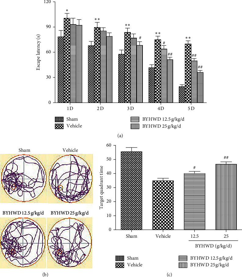
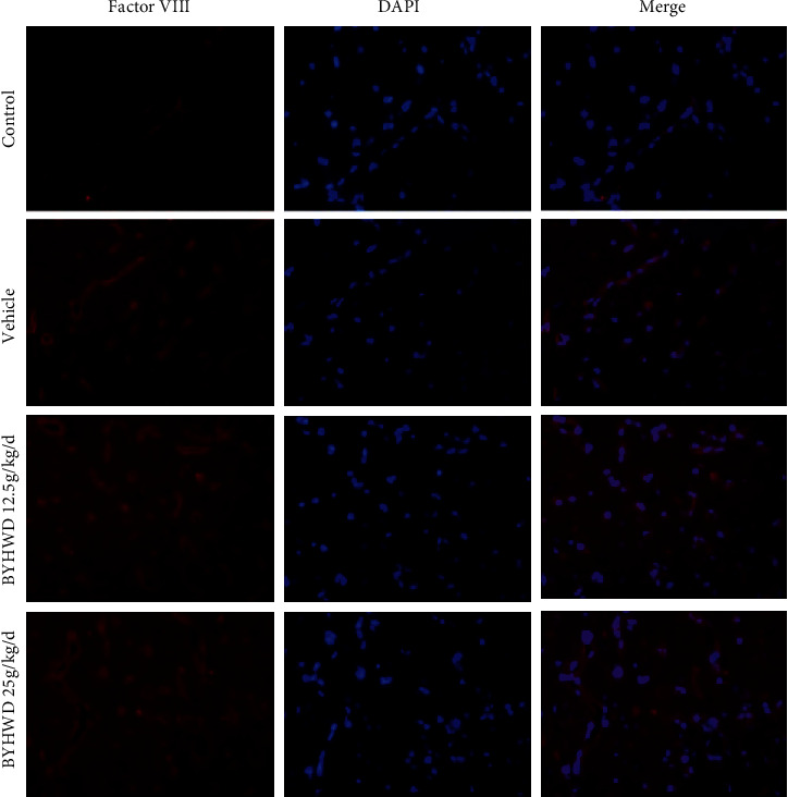
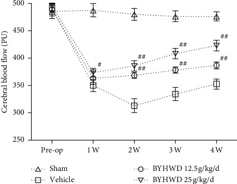
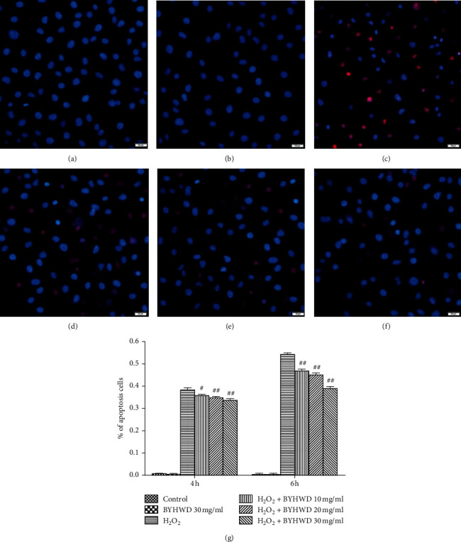
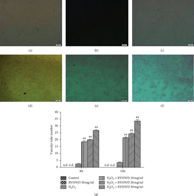
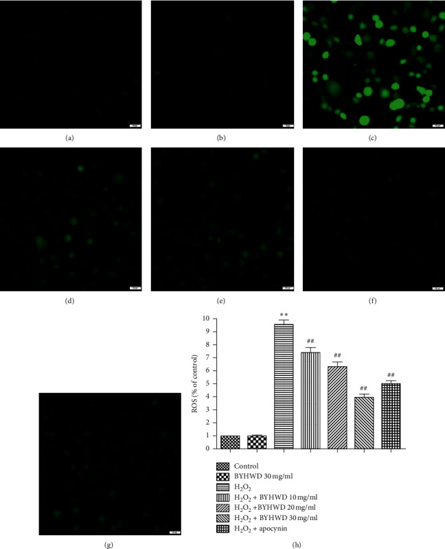
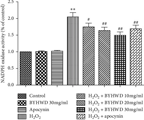
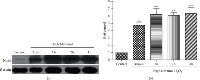
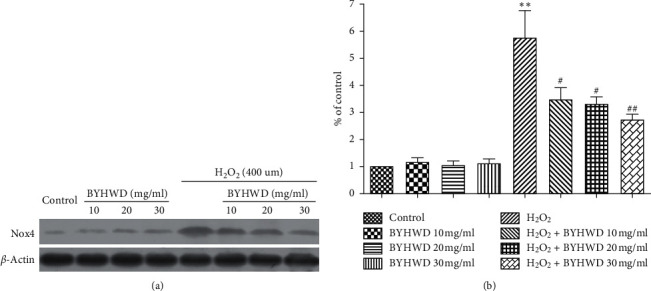
Methods: The effects of BYHWD on neurological function were screened by measuring neurological deficits, spatial memory function, and angiogenesis (by microvascular density (MVD) and cerebral blood flow (CBF)) after CI/R injury in middle cerebral artery occlusion (MCAO) in vivo in rats. In vitro, we examined the angiogenic roles and mechanisms of action of BYHWD in an H2O2-induced oxidative stress HUVECs model by measuring cell viability, apoptosis, vascular tube formation, intracellular ROS generation, NADPH oxidase (Nox) activity, and Nox4 protein expression.

Results: BYHWD significantly improved neurological function, including neurological deficits and spatial learning and memory, and significantly increased MVD and CBF in the ischemic penumbra after CI/R injury in rats. BYHWD significantly increased cell viability, inhibited apoptosis, induced vascular tube formation, decreased intracellular ROS generation, and reduced Nox activity and Nox4 protein expression in H2O2-treated HUVECs in a dose-dependent manner.

Conclusions: Our study demonstrates that BYHWD promotes neurological function recovery and increases angiogenesis. BYHWD exerts angiogenic effects against cerebral ischemic injury through the downregulation of Nox4, which results in the reduction of ROS generation.


Conflict of interest statement

All authors declare that they have no conflicts of interest.

Figures

Figure 1
Figure 1
Effects of BYHWD on neurological deficit after focal cerebral ischemia in rats. The neurological deficit for each group was measured at 1, 2, 3, and 4 w after focal cerebral ischemia. #P < 0.05 and ##P < 0.01 versus vehicle group on the same day (n = 10).
Figure 2
Figure 2
Effects of BYHWD in spatial acquisition trials and probe trials of Morris water maze after focal cerebral ischemia in rats. (a) Latency for reaching the hidden platform during 5 consecutive days of spatial acquisition training. (b) Representative search patterns during the probe transfer trial. (c) Time spent in the quadrant where the platform had previously been located during the probe transfer trial. #P < 0.05 and ##P < 0.01 versus vehicle group on the same day (n = 10).
Figure 3
Figure 3
Effects of BYHWD on microvessel density (MVD) after focal cerebral ischemia in rats. Representative microphotographs of factor VIII-positive MVD for each group at 4 w after focal cerebral ischemia (n = 6). Scale bar, 50 μm.
Figure 4
Figure 4
Effect of BYHWD on cerebral blood flow (CBF) after focal cerebral ischemia in rats. CBF for each group before the operation, and 1, 2, 3, and 4 w after focal cerebral ischemia. #P < 0.05 and ##P < 0.01 versus vehicle group on the same day (n = 10).
Figure 5
Figure 5
Effects of BYHWD on H2O2-induced cytotoxicity in HUVECs. Cell viability in HUVECs preincubated with BYHWD (10, 20, or 30 mg/ml) for 6 h followed by H2O2 for 4 or 6 h #P < 0.05 and ##P < 0.01 versus H2O2 group at the same time.
Figure 6
Figure 6
Effects of BYHWD on H2O2-induced cell apoptosis in HUVECs. Cells were double-stained with cell membrane-permeable (Hoechst 33342; blue) and impermeable (PI; red) DNA labeling fluorochromes. (a) Vehicle medium (control) for 6 h. (b) Preincubation with BYHWD (30 mg/ml) for 6 h. (c) H2O2 (400 μM) for 6 h. (d) Preincubation with BYHWD (10 mg/ml) for 6 h followed by H2O2 for 6 h. (e) Preincubation with BYHWD (20 mg/ml) for 6 h followed by H2O2 for 6 h. (f) Preincubation with BYHWD (30 mg/ml) for 6 h followed by H2O2 for 6 h. (g) The percentage of apoptotic cells for each group and time point. #P < 0.05 and ##P < 0.01 versus the H2O2 group at the same time. Scale bars, 50 μm.
Figure 7
Figure 7
Effects of BYHWD on H2O2-induced vascular tube formation in HUVECs. (a) Vehicle medium (control) for 10 h. (b) Preincubation with BYHWD (30 mg/ml) for 6 h. (c) H2O2 (400 μM) for 10 h. (d) Preincubation with BYHWD (10 mg/ml) for 6 h followed by H2O2 for 10 h. (e) Preincubation with BYHWD (20 mg/ml) for 6 h followed by H2O2 for 10 h. (f) Preincubation with BYHWD (30 mg/ml) for 6 h followed by H2O2 for 10 h. (g) Numbers of vascular tubes in HUVECs cultures for each group and time point. ##P < 0.01 versus the H2O2 group at the same time. Scale bars, 100 μm.
Figure 8
Figure 8
Effects of BYHWD on H2O2-induced intracellular ROS in HUVECs. Cells were stained with DCFH-DA. (a) Vehicle medium (control) for 6 h. (b) Preincubation with BYHWD (30 mg/ml) for 6 h. (c) H2O2 (400 μM) for 6 h. (d) Preincubation with BYHWD (10 mg/ml) for 6 h followed by H2O2 for 6 h. (e) Preincubation with BYHWD (20 mg/ml) for 6 h followed by H2O2 for 6 h. (f) Preincubation with BYHWD (30 mg/ml) for 6 h followed by H2O2 for 6 h. (g) Preincubation with apocynin (200 μM) for 1 h followed by H2O2 for 6 h. (h) Intracellular ROS level in HUVECs for each group. ∗∗P < 0.01 versus the control group. ##P < 0.01 versus the H2O2 group. Scale bars, 50 μm.
Figure 9
Figure 9
Effects of BYHWD on H2O2-induced NADPH oxidase activity in HUVECs. NADPH oxidase activity in HUVECs preincubated with BYHWD (10, 20, or 30 mg/ml) for 6 h or apocynin (200 μM) for 1 h followed by H2O2 for 2 h ∗∗P < 0.01 versus the control group. #P < 0.05 and ##P < 0.01 versus the H2O2 group.
Figure 10
Figure 10
Time course of the effects of H2O2 on Nox4 protein expression in HUVECs. (a) Western blot of Nox4 protein expression in HUVECs treated with H2O2 (400 μmol/L) for various lengths of time (30 min or 1, 2, or 4 h). (b) Quantification of Nox4 protein expression in HUVECs for each group. ∗∗P < 0.01 versus the control group.
Figure 11
Figure 11
Effects of BYHWD on H2O2-induced Nox4 protein expression in HUVECs. (a) Western blot of Nox4 protein expression in HUVECs preincubated with BYHWD (10, 20, or 30 mg/ml) for 6 h followed by H2O2 for 2 h. (b) Quantification of Nox4 protein expression in HUVECs for each group. ∗∗P < 0.01 versus the control group. #P < 0.05 and ##P < 0.01 versus the H2O2 group.

References

    1. GBD 2016 Causes of Death Collaborators. Global, regional, and national age-sex specific mortality for 264 causes of death, 1980–2016: a systematic analysis for the Global Burden of Disease Study 2016. The Lancet. 2017;390:1151–1210. - PMC - PubMed
    1. Liu J., Wang Y., Akamatsu Y., et al. Vascular remodeling after ischemic stroke: mechanisms and therapeutic potentials. Progress in Neurobiology. 2014;115:138–156. doi: 10.1016/j.pneurobio.2013.11.004. - DOI - PMC - PubMed
    1. Kahles T., Luedike P., Endres M., et al. NADPH oxidase plays a central role in blood-brain barrier damage in experimental stroke. Stroke. 2007;38(11):3000–3006. doi: 10.1161/strokeaha.107.489765. - DOI - PubMed
    1. Hu Y., Li R., Yang H., Luo H., Chen Z. Sirtuin 6 is essential for sodium sulfidemediated cytoprotective effect in ischemia/reperfusion-stimulated brain endothelial cells. Journal of Stroke and Cerebrovascular Diseases. 2015;24(3):601–609. doi: 10.1016/j.jstrokecerebrovasdis.2014.10.006. - DOI - PubMed
    1. Jung Y. S., Lee S. W., Park J. H., Seo H. B., Choi B. T., Shin H. K. Electroacupuncture preconditionin reduces ROS generation with Nox4 down-regulation and ameliorates blood-brain barrier disruption after ischemic stroke. Journal of Biomedical Science. 2016;23:p. 32. doi: 10.1186/s12929-016-0249-0. - DOI - PMC - PubMed

LinkOut - more resources