. 2020 Oct:34:100898.
doi: 10.1016/j.nantod.2020.100898. Epub 2020 Jun 20.

Overcoming negatively charged tissue barriers: Drug delivery using cationic peptides and proteins

Affiliations

Overcoming negatively charged tissue barriers: Drug delivery using cationic peptides and proteins

Armin Vedadghavami et al. Nano Today. 2020 Oct.

Abstract

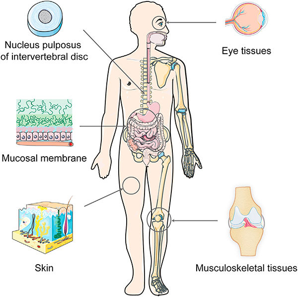
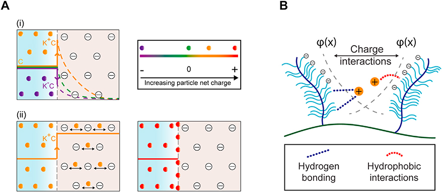
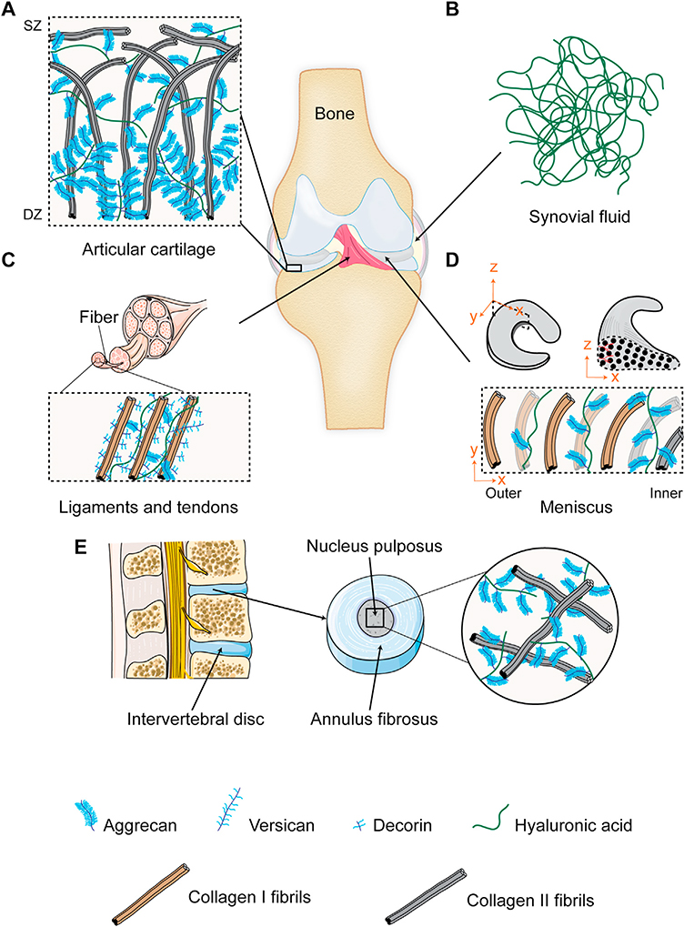
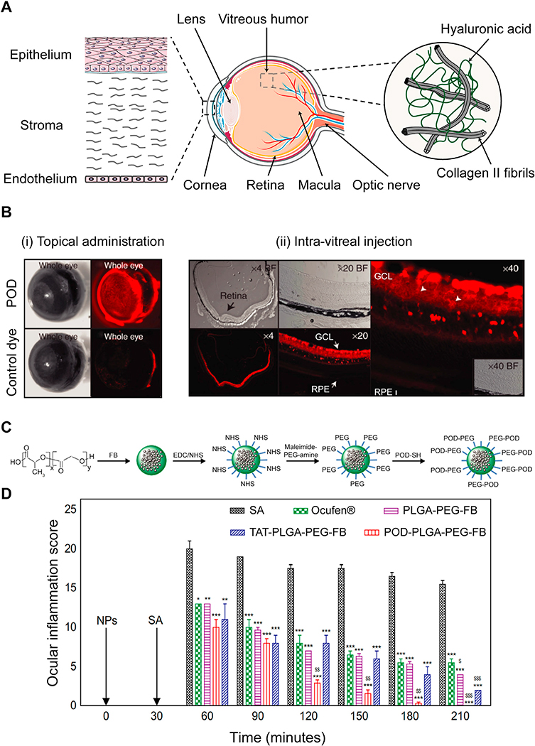
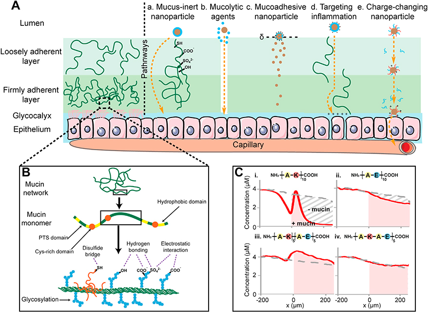
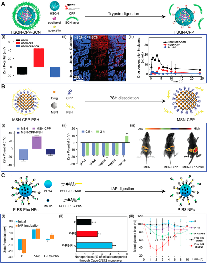
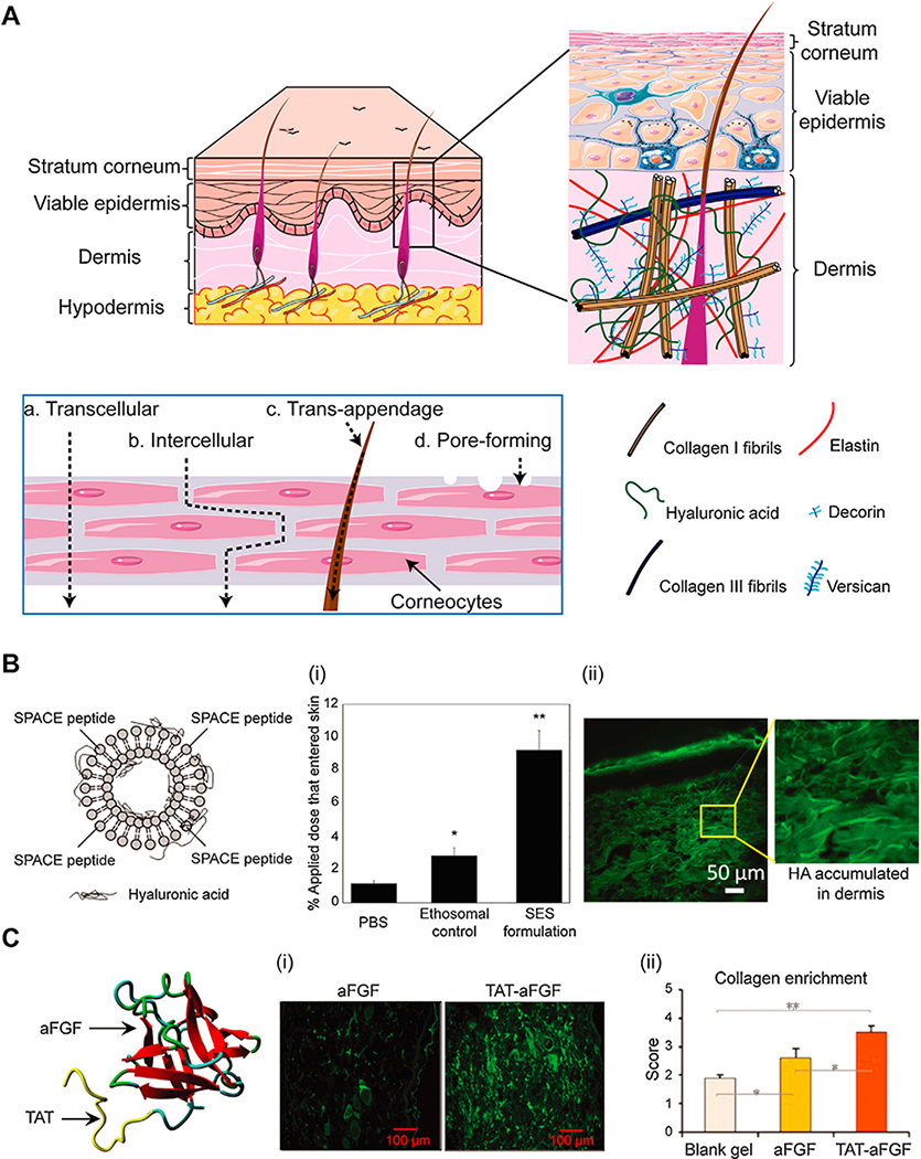
Negatively charged tissues are ubiquitous in the human body and are associated with a number of common diseases yet remain an outstanding challenge for targeted drug delivery. While the anionic proteoglycans are critical for tissue structure and function, they make tissue matrix dense, conferring a high negative fixed charge density (FCD) that makes drug penetration through the tissue deep zones and drug delivery to resident cells extremely challenging. The high negative FCD of these tissues is now being utilized by taking advantage of electrostatic interactions to create positively charged multi-stage delivery methods that can sequentially penetrate through the full thickness of tissues, create a drug depot and target cells. After decades of work on attempting delivery using strong binding interactions, significant advances have recently been made using weak and reversible electrostatic interactions, a characteristic now considered essential to drug penetration and retention in negatively charged tissues. Here we discuss these advances using examples of negatively charged tissues (cartilage, meniscus, tendons and ligaments, nucleus pulposus, vitreous of eye, mucin, skin), and delve into how each of their structures, tissue matrix compositions and high negative FCDs create barriers to drug entry and explore how charge interactions are being used to overcome these barriers. We review work on tissue targeting cationic peptide and protein-based drug delivery, compare and contrast drug delivery designs, and also present examples of technologies that are entering clinical trials. We also present strategies on further enhancing drug retention within diseased tissues of lower FCD by using synergistic effects of short-range binding interactions like hydrophobic and H-bonds that stabilize long-range charge interactions. As electrostatic interactions are incorporated into design of drug delivery materials and used as a strategy to create properties that are reversible, tunable and dynamic, bio-electroceuticals are becoming an exciting new direction of research and clinical work.

Keywords: Cationic drug carriers; Cationic protein drug carriers; Cell penetrating peptides; Electro-diffusive transport; Electrostatic charge interactions; Negatively charged tissues; Targeted drug delivery.


Conflict of interest statement

Declaration of Competing Interest The authors declare no conflict of interest.

Figures

Fig. 1.
Fig. 1.
Negatively charged tissues in the human body remain an outstanding challenge in the field of targeted drug delivery. Examples include musculoskeletal joints(shoulder, elbow, knee, hand and ankle) comprising of tissues like articular cartilage, synovial joint, ligaments, tendons and menisci; nucleus pulposus of the intervertebral disc; eye tissues like the cornea and vitreous humor; mucosal membrane; and skin comprising of multiple layers of negatively charged cells, collagen, proteoglycans and elastin. This high negative FCD can be used to enhance intra-tissue transport, uptake and binding of locally injected drugs or their carriers via electrostatic interactions by modifying them to contain optimally charged cationic domains.
Fig. 2.
Fig. 2.
Electro-diffusive transport of cationic carriers in negatively charged tissues. A. (i) Concentration of positively charged carriers partitions upward from C to K+C at tissue interface enabling faster diffusion rates compared to their neutral counterpart. Conversely, concentration of negatively charged carriers partitions downward from C to KC due to electrostatic repulsion resulting in slower intra-tissue diffusion rates. (ii) Optimally charged cationic carriers can penetrate through the full thickness of tissues owing to weak-reversible binding nature of charge interactions while carriers with too high positive charge get stuck within the tissue surface due to strong binding interactions. B. Incorporation of short-range effects such as from hydrophobic or hydrogen bonds can synergistically stabilize long-range charge-based binding of cationic carriers with their intra-tissue negatively charged binding sites. φ(x) denotes the electric potential exerted by fixed negatively charged groups as a function of distance, x inside the tissue.
Fig. 3.
Fig. 3.
Musculoskeletal joint tissues. A. Articular cartilage is comprised of a network of collagen II and a high density of aggrecans. Collagen fibrils are aligned parallel to the surface in the superficial zone (SZ) while arranged perpendicularly in the cartilage deep zone (DZ); aggrecan density increases from SZ to DZ. B. Synovial fluid is comprised of high molecular weight hyaluronic acid meshwork. C. Ligaments and tendons are comprised of linearly aligned collagen I fibrils containing decorin, aggrecans and versicans. D. Meniscus is comprised of circumferentially aligned collagen fibrils, mainly type I in the outer two-third region and a combination of types I and II in the inner region. Aggrecan density is highest in the inner region and decreases towards the outer region. E. Intervertebral disc is comprised ofgelatinous nucleus pulposus containing a random network of collagen II and proteoglycans.
Fig.4.
Fig.4.
A. (i) Full-thickness penetration of positively charged Avidin in rabbit femoral cartilage within 24 h of IA injection. Adapted from [53]. (ii) IHC staining shows presence of Avidin in rabbit cartilage even at 3 weeks after ACLT surgery. (iii) μCT of femoral condyles show significantly suppressed osteophyte volume with Av-Dex compared to free Dex. Adapted from ref [16]. Printed with permission from AO Research Institute Davos. B. (i) Multi-arm Avidin nanoconstruct conjugated to Dex (mAv-Dex) using ester linkers. (ii) Safranin-O/fast green staining of cartilage explants shows mAv-Dex significantly suppressed IL-1 induced GAG loss compared to free Dex over 16 days. Adapted from ref [55]. Printed with permission from Elsevier. C. Cationic PAMAM dendrimer for IGF-1 delivery. (i) 3D reconstruction of multiphoton microscopy of rat cartilage. Gen 6 45 % PEG-IGF-1 PAMAM dendrimer shows superior intra-cartilage penetration and retention compared to free IGF-1 at 6 days post IA injection in rats. Aggrecans, collagen and IGF-1 are marked in red, grey and blue, respectively. (ii) Toluidine blue/fast green staining of rat cartilage show PEG-IGF-1 dendrimer suppressed GAG loss greaterthan free IGF-1 after 4 weeks of ACLT surgery. Adapted from ref [35]. Printed with permission from The American Association for the Advancement of Science. D. (i) DOTAM nanoparticles with collagen II targeting peptide (TP) and amines. (ii) Fluorescent images show increased DOTAM-TP retention in mice knees 48 h post IA injection compared to control probe. (iii) Histological analysis of mice cartilage show intra-cartilage localization and retention of DOTAM-TP. Adapted from ref [63]. Printed with permission from American Chemical Society.
Fig. 5.
Fig. 5.
A. Structure of ocular tissue in the anterior (cornea and the lens) and posterior (vitreous humor, retina and macula) regions. Barriers to local drug delivery include negatively charged corneal epithelium layer and vitreous humor comprising of collagen II and hyaluronic acid that prevent diffusion of drugs and their carriers to the posterior regions. B. Transport of POD in ocular tissues upon (i) topical administration and (ii) intra-vitreal injection. (i) POD was uptaken by external ocular tissues within 45 minutes upon topical administration in mice eyes while the control free dye only weakly stained the eye. (ii) Intra-vitreal injection of POD transduced 85 % of neural retina within 2 h. RPE and GCL represent retinal pigment epithelium and ganglion cell layer, respectively. Adapted from ref [137]. Printed with permission from Elsevier. C. Synthesis of POD-PLGA-PEG-FB nanoparticles. D. Effectiveness of POD-PLGA-PEG-FB in prevention of sodium arachidonate (SA) induced ocular inflammation in rabbit eyes (*p < 0.05, **p < 0.01, and ***p < 0.001 vs inflammation induced by SA.$p< 0.05, $ $p< 0.01, and $ $ $p < 0.001 vs anti-inflammatory effect of Ocufen®). Adapted from ref [140]. Printed with permission under Creative Commons Attribution License.
Fig. 6.
Fig. 6.
A. Characteristics of the mucus barrier coating epithelium and strategies used for promoting mucus penetration (a. Mucus-inert nanoparticle b. Mucolytic agents c. Mucoadhesive nanoparticle d. Targeting inflammation e. Charge-changing nanoparticle). B. Architecture of the mucus network and structure of the heavily glycosylated mucin monomer. C. Charge-based transport behavior of short length peptides of varying net charge and spatial distribution in mucin gel, i. Cationic peptide, ii. Anionic peptide, iii. Block peptide and iv. Alternate peptide. Adapted from ref [45]. Printed with permission from Elsevier.
Fig. 7.
Fig. 7.
Charge-reversal nanoparticle systems developed for both effective mucus penetration and GI epithelium targeting. A. Quantum dots doped hollow silica nanoparticles(HSQN) modified by CPPs with a cleavable outermost shell of hydrophilic succinylated casein (SCN). (i) Zeta potential, (ii) Confocal laser scanning microscopy images of rat intestine 2 h post oral administration (blue showing cell nuclei and red showing presence of nanoparticles). (iii) Drug levels in plasma after oral administration of HSQN-CPP-SCN. Adapted from ref [172]. Printed with permission from Elsevier. B. Mesoporous silica nanoparticles (MSN) with an interlayer of CPP and an outermost shell of thiolated polymer (PSH). (i) Zeta potential, (ii) Enzymatic and pH stability of MSN-CPP-PSH showing specific degradation of PSH shell in presence of mucus that reverses its net charge. (iii) In vivo bio-distribution of MSN-CPP-PSH after oral administration in mice. Adapted from ref [173]. Printed with permission from Elsevier. C. PLGA nanoparticles coated with R8 and anionic phosphoserine (Pho), which dissociates upon digestion with intestinal alkaline phosphatase (IAP) leading to a net positive surface charge. (i) Zeta potential, (ii) Enhanced in-vitro transepithelial transport of P-R8-Pho (iii) Suppression of blood glucose levels after oral administration of insulin-loaded P-R8-Phonanoparticles in diabetic rats. Adapted from ref [174]. Printed with permission from American Chemical Society.
Fig. 8.
Fig. 8.
A. Different layers of skin (stratum corneum, viable epidermis, dermis and hypodermis). Sublayers of viable epidermis and dermis are comprised of a collagen network, elastin, versican and HA. Different routes of transdermal drug delivery (a. Transcellular, b. Intercellular, c. Trans-appendage and d. Pore-forming). B. SPACE peptide conjugated to phospholipids designed as an ethosomal carrier system (SES) to deliver hyaluronic acid (HA). (i) Percent penetration oftopically applied dose into total skin. (ii) Confocal image of dermis showing SES can penetrate through the full skin to deliver HA. Adapted from ref[196]. Printed with permission from Elsevier. C. Conjugation of TAT to aFGF to improve its transdermal delivery efficiency. (i) Immunofluorescence of aFGF or TAT-aFGF accumulated in dermal and subcutaneous tissues. (ii) Semi-quantitative scoring showing greater collagen enrichment in rat skin injury model with TAT-aFGF. Adapted from ref[199]. Printed with permission under Creative Commons Attribution License.

Similar articles

Cited by

References

    1. Zhang Y,Jordan JM, Epidemiology of osteoarthritis, Clin. Geriatr. Med. 26 (2010) 355–369, 10.1016/jxger.2010.03.001. - DOI - PMC - PubMed
    1. Roddy E, Choi HK, Epidemiology of gout, Rheum. Dis. Clin. North Am. 40 (2014) 155–175, 10.1016/j.rdc.2014.01.001. - DOI - PMC - PubMed
    1. Freburger JK, Holmes GM, Agans RP, Jackman AM,Darter JD, Wallace AS, Castel LD, Kalsbeek WD, Carey TS, The rising prevalence of chronic low back pain, Arch. Intern. Med. 169 (2009) 251–258, 10.1001/archinternmed.2008.543. - DOI - PMC - PubMed
    1. Ananthakrishnan AN, Epidemiology and risk factors for IBD, Nat. Rev. Gastroenterol. Hepatol. 12(2015) 205–217, 10.1038/nrgastro.2015.34. - DOI - PubMed
    1. Wong WL, Su X, Li X, Cheung CMG, Klein R, Cheng CY, Wong TY, Global prevalence ofage-related macular degeneration and disease burden projection for 2020 and 2040: a systematic review and meta-analysis, Lancet Glob. Heal. 2 (2014), 10.1016/S2214-109X(13)70145-1. - DOI - PubMed

LinkOut - more resources