. 2020 Jul 29:2020:2405135.
doi: 10.1155/2020/2405135. eCollection 2020.

Peroxiredoxin-1 Overexpression Attenuates Doxorubicin-Induced Cardiotoxicity by Inhibiting Oxidative Stress and Cardiomyocyte Apoptosis

Affiliations

Peroxiredoxin-1 Overexpression Attenuates Doxorubicin-Induced Cardiotoxicity by Inhibiting Oxidative Stress and Cardiomyocyte Apoptosis

Lai Jiang et al. Oxid Med Cell Longev. .

Abstract

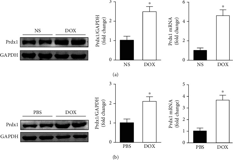
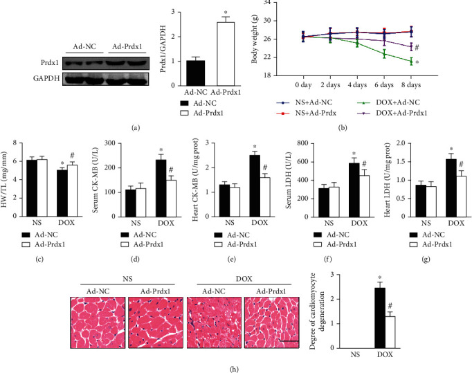
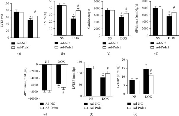
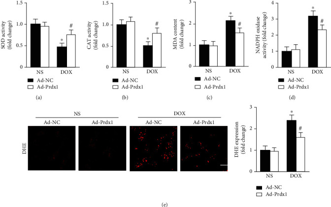
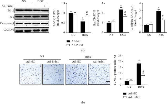
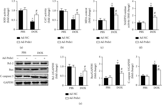
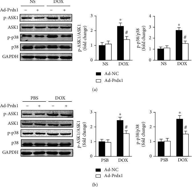
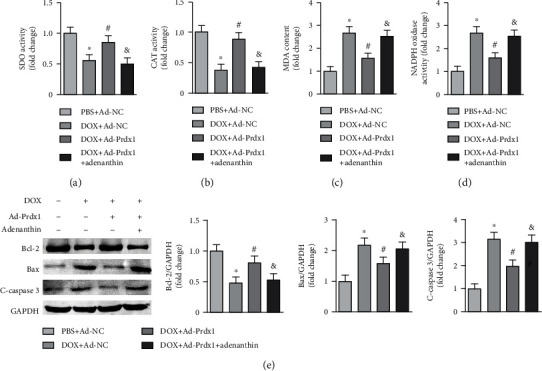
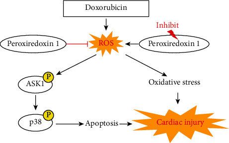
Background. Previous research has shown that peroxiredoxin 1 (Prdx1) is an important modulator of physiological and pathophysiological cardiovascular events. This study is aimed at investigating the role and underlying mechanism of Prdx1 in doxorubicin- (DOX-) induced cardiotoxicity. Cardiac-specific expression of Prdx1 was induced in mice, and the mice received a single dose of DOX (15 mg/kg) to generate cardiotoxicity. First, our study demonstrated that Prdx1 expression was upregulated in the heart and in cardiomyocytes after DOX treatment. Second, we provided direct evidence that Prdx1 overexpression ameliorated DOX-induced cardiotoxicity by attenuating oxidative stress and cardiomyocyte apoptosis. Mechanistically, we found that DOX treatment increased the phosphorylation level of apoptosis signal-regulating kinase-1 (ASK1) and the downstream protein p38 in the heart and in cardiomyocytes, and these effects were decreased by Prdx1 overexpression. In contrast, inhibiting Prdx1 promoted DOX-induced cardiac injury via the ASK1/p38 pathway. These results suggest that Prdx1 may be an effective therapeutic option to prevent DOX-induced cardiotoxicity.


Conflict of interest statement

No conflicts of interest are declared by the authors.

Figures

Figure 1
Figure 1
Prdx1 expression is increased in the heart and in cardiomyocytes after DOX treatment. (a) The protein and mRNA expression of Prdx1 in the heart 8 days after DOX treatment (n = 4, P < 0.05 compared with the NS group). (b) The protein and mRNA expression of Prdx1 in cardiomyocytes treated with DOX (n = 4, P < 0.05 compared with the PBS group).
Figure 2
Figure 2
Prdx1 overexpression protected against cardiac injury induced by DOX. (a) The protein levels of Prdx1 four weeks after AAV9-Prdx1 injection in mice (n = 4). (b, c) The results of body weight and HW/TL ratio measurements in mice (n = 6). (d–g) Biochemical determination of CK-MB and LDH levels in the heart and serum for the indicated groups (n = 6). (h) HE staining shows the pathological structure of the heart in mice (n = 5). P < 0.05 compared with the NS group; #P < 0.05 compared with the DOX group.
Figure 3
Figure 3
Prdx1 overexpression attenuated cardiac dysfunction induced by DOX. (a, b) Echocardiographic parameters for each group (n = 8). (c–g) Hemodynamic parameters for each group (n = 6). P < 0.05 compared with the NS group; #P < 0.05 compared with the DOX group.
Figure 4
Figure 4
Prdx1 overexpression attenuated oxidative stress induced by DOX. (a–d) Quantitative results for SOD, CAT, NADPH oxidase activity, and MDA content in the hearts of mice (n = 6). (e) Representative DHE staining images of the heart (n = 5). P < 0.05 compared with the NS group; #P < 0.05 compared with the DOX group.
Figure 5
Figure 5
Prdx1 overexpression attenuated cardiomyocyte apoptosis induced by DOX. (a) Western blots showing the Bcl-2, Bax, and c-caspase3 levels in the hearts of mice (n = 4). (b) Representative TUNEL staining images of the heart (n = 5). P < 0.05 compared with the NS group; #P < 0.05 compared with the DOX group.
Figure 6
Figure 6
Prdx1 overexpression relieves DOX-induced cardiotoxicity in vitro. (a–d) Quantitative results for SOD, CAT, NADPH oxidase activity, and MDA content in each group (n = 6). (e) Western blots showing the Bcl-2, Bax, and c-caspase3 levels in each group (n = 4). P < 0.05 compared with the PBS group; #P < 0.05 compared with the DOX group.
Figure 7
Figure 7
Prdx1 overexpression inhibits ASK1/p38 pathway activation. (a) Western blots showing the p-ASK1, ASK1, p-p38, and p38 levels in the hearts of mice (n = 4). (b) Western blots showing the p-ASK1, ASK1, p-p38, and p38 levels in NVRMs (n = 4). P < 0.05 compared with the NS or PBS group; #P < 0.05 compared with the DOX group.
Figure 8
Figure 8
Prdx1 inhibition exacerbated DOX-induced cardiotoxicity. (a–d) Quantitative results for SOD, CAT, NADPH oxidase activity, and MDA content in each group (n = 6). (e) Western blots showing the Bcl-2, Bax, and c-caspase3 levels in each group (n = 4). P < 0.05 compared with the PBS+Ad-NC group; #P < 0.05 compared with the DOX+Ad-NC group; &P < 0.05 compared with the DOX+Ad-Prdx1 group.
Figure 9
Figure 9
Peroxiredoxin-1 overexpression attenuates doxorubicin-induced cardiotoxicity by inhibiting oxidative stress and cardiomyocyte apoptosis.

Similar articles

Cited by

References

    1. Campia U., Moslehi J. J., Amiri-Kordestani L., et al. Cardio-oncology: vascular and metabolic perspectives: a scientific statement from the American Heart Association. Circulation. 2019;139(13):e579–e602. doi: 10.1161/CIR.0000000000000641. - DOI - PMC - PubMed
    1. Herrmann J. Adverse cardiac effects of cancer therapies: cardiotoxicity and arrhythmia. Nature Reviews Cardiology. 2020;17(8):474–502. doi: 10.1038/s41569-020-0348-1. - DOI - PMC - PubMed
    1. Chen Y., Huang T., Shi W., Fang J., Deng H., Cui G. Potential targets for intervention against doxorubicin-induced cardiotoxicity based on genetic studies: a systematic review of the literature. Journal of Molecular and Cellular Cardiology. 2020;138:88–98. doi: 10.1016/j.yjmcc.2019.11.150. - DOI - PubMed
    1. Gorini S., De Angelis A., Berrino L., Malara N., Rosano G., Ferraro E. Chemotherapeutic drugs and mitochondrial dysfunction: focus on doxorubicin, trastuzumab, and sunitinib. Oxidative Medicine and Cellular Longevity. 2018;2018:15. doi: 10.1155/2018/7582730.7582730 - DOI - PMC - PubMed
    1. Carvalho F. S., Burgeiro A., Garcia R., Moreno A. J., Carvalho R. A., Oliveira P. J. Doxorubicin-induced cardiotoxicity: from bioenergetic failure and cell death to cardiomyopathy. Medicinal Research Reviews. 2014;34(1):106–135. doi: 10.1002/med.21280. - DOI - PubMed

LinkOut - more resources