. 2020 Aug 5;5(1):73.
doi: 10.1038/s41541-020-00220-4. eCollection 2020.

A replication-defective Japanese encephalitis virus (JEV) vaccine candidate with NS1 deletion confers dual protection against JEV and West Nile virus in mice

Affiliations

A replication-defective Japanese encephalitis virus (JEV) vaccine candidate with NS1 deletion confers dual protection against JEV and West Nile virus in mice

Na Li et al. NPJ Vaccines. .

Abstract

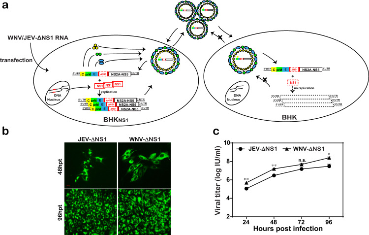
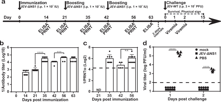
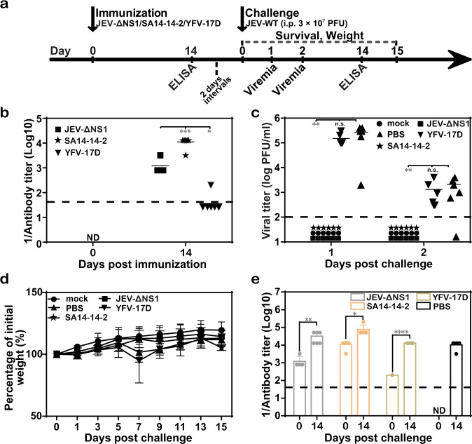
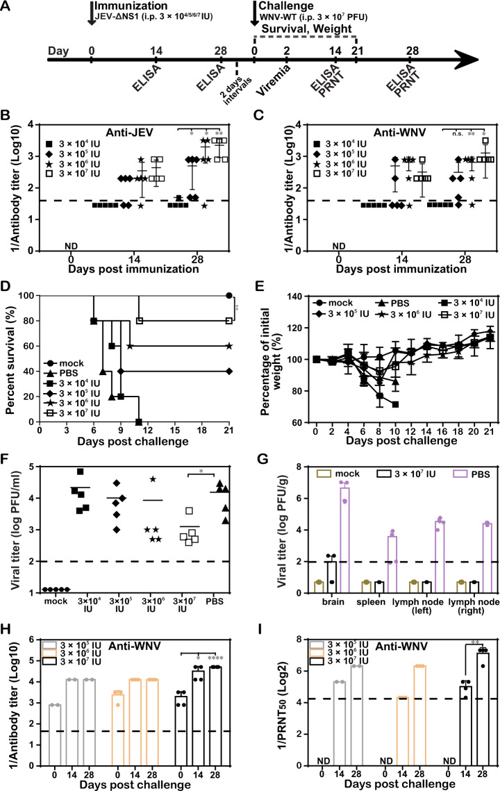
In our previous study, we have demonstrated in the context of WNV-ΔNS1 vaccine (a replication-defective West Nile virus (WNV) lacking NS1) that the NS1 trans-complementation system may offer a promising platform for the development of safe and efficient flavivirus vaccines only requiring one dose. Here, we produced high titer (107 IU/ml) replication-defective Japanese encephalitis virus (JEV) with NS1 deletion (JEV-ΔNS1) in the BHK-21 cell line stably expressing NS1 (BHKNS1) using the same strategy. JEV-ΔNS1 appeared safe with a remarkable genetic stability and high degrees of attenuation of in vivo neuroinvasiveness and neurovirulence. Meanwhile, it was demonstrated to be highly immunogenic in mice after a single dose, providing similar degrees of protection to SA14-14-2 vaccine (a most widely used live attenuated JEV vaccine), with healthy condition, undetectable viremia and gradually rising body weight. Importantly, we also found JEV-ΔNS1 induced robust cross-protective immune responses against the challenge of heterologous West Nile virus (WNV), another important member in the same JEV serocomplex, accounting for up to 80% survival rate following a single dose of immunization relative to mock-vaccinated mice. These results not only support the identification of the NS1-deleted flavivirus vaccines with a satisfied balance between safety and efficacy, but also demonstrate the potential of the JEV-ΔNS1 as an alternative vaccine candidate against both JEV and WNV challenge.

Keywords: Live attenuated vaccines; Vaccines.


Conflict of interest statement

Competing interestsThe authors declare no competing interests.

Figures

Fig. 1
Fig. 1. High replication efficiency of JEV-ΔNS1 in BHKNS1 cell line.
a Schematic diagram of the generation and replication of JEV-ΔNS1 particles in cells. JEV-ΔNS1 (with a deletion of the residues 4–298 in NS1 coding sequence) replicates efficiently in the BHK-21 cell line stably expressing WNV-NS1 protein (BHKNS1), while undergoes a single round of entry, release and viral RNA translation in the normal cells. b IFA detection of JEV-ΔNS1 and WNV-ΔNS1 in BHKNS1 cells post transfection. Equal amounts of JEV-ΔNS1 and WNV-ΔNS1 RNAs were transfected into BHKNS1 cells. IFA analysis using 4G2 monoclonal antibody was performed at the indicated time points. The length of the scale bar (displayed in a red line segment) represents 20 μm. c Comparison of growth kinetics of JEV-ΔNS1 and WNV-ΔNS1. BHKNS1 cells were infected with JEV-ΔNS1 and WNV-ΔNS1 virus at an MOI of 0.1. The supernatants were harvested at the indicated time points and viral titers were determined by IFA on BHKNS1 cells as described in “Methods”. Two independent experiments were performed in triplicate. Data represent the mean ± standard deviation (SD) of the triplicate measurements in a representative experiment. Statistical analysis was performed with unpaired t test and the asterisks denote statistical differences between the indicated groups. *p < 0.05; **p < 0.01; n.s. no statistical difference.
Fig. 2
Fig. 2. Characterization of JEV-ΔNS1.
a Comparison of growth kinetics between WT JEV (JEV-WT) and JEV-ΔNS1 virus in BHKNS1 cells. BHKNS1 cells were infected with either JEV-WT or JEV-ΔNS1 virus at an MOI of 0.1. The supernatants were harvested at the indicated time points and viral titers were determined as described above. Data represent the mean ± standard deviation (SD) of the triplicate measurements in a representative experiment. Statistical analysis was performed with unpaired t test and the asterisks denote statistical differences between the indicated groups. **p < 0.01; n.s. no statistical difference. b Western blotting analysis of purified JEV-WT and JEV-ΔNS1 viral particles. Following concentration and purification through PEG8000 precipitation and ultracentrifugation, equal amounts of purified WT and ΔNS1 viruses were subjected to Western blotting analysis using the specific anti-E monoclonal antibody or anti-C polyclonal antibody. All blots derived from the same experiment and were processed in parallel. c ELISA for the detection of the antibodies against both JEV and JEV-ΔNS1. Two independent experiments were performed in triplicate. Data represent the mean ± standard deviation (SD) of the triplicate measurements in a representative experiment. Statistical analysis was performed with unpaired t test. n.s. no statistical difference.
Fig. 3
Fig. 3. Stability of JEV-ΔNS1.
a IFA analysis of JEV-ΔNS1 virus at different passages using 4G2 monoclonal antibody. The JEV-ΔNS1 virus generated through transfection of BHKNS1 cells with the transcribed JEV-ΔNS1 genomic RNA was designated as P0 virus. Three JEV-ΔNS1 virus stocks (A, B and C) were blind passaged independently for 15 rounds (P0−P15) in BHKNS1 cells. The viruses at P0 and P15 were used to infect BHKNS1 cells and naïve BHK-21 cells. JEV-WT was used as a positive control. The length of the scale bar (displayed in a red line segment) represents 20 μm. b RT-PCR analysis of the expression of NS1 gene in BHKNS1 cells infected with P0 or P15 JEV-ΔNS1 viruses. The uncropped and unprocessed gel including markers was displayed in Supplementary Fig. 2. c Sequence chromatograms of RT-PCR products containing NS1 fragment amplified from P15 JEV-ΔNS1 virus.
Fig. 4
Fig. 4. JEV-ΔNS1 is highly safe in mice.
a, b Neuroinvasiveness tests for WT and JEV-ΔNS1 viruses in mice. Four-week-old C57BL/6 female mice (n = 5 for each group) were i.p. inoculated with 106 and 107 IU of WT JEV or 107 IU of JEV-ΔNS1. The mortality (a) and weight loss (b) were monitored for 21 days. c, d Neurovirulence tests for WT and JEV-ΔNS1 viruses in mice. Three-week-old ICR female mice (n = 5 for each group) were i.c. inoculated with 10 IU of WT JEV or 7.5 × 106 IU of JEV-ΔNS1. The mortality (c) and weight loss (d) were monitored for 21 days. Kaplan−Meier survival curves were analyzed by the log-rank test and the asterisks denote statistical differences between the indicated groups. *p < 0.05; ***p < 0.001. Two independent experiments were performed, and data from one experiment are presented.
Fig. 5
Fig. 5. Protection of JEV-ΔNS1 in mice against JEV challenge.
a Schematic diagram of animal experiment design and schedule. Four-week-old C57BL/6 female mice (n = 10 for each group) were i.p. immunized three times at 3-week intervals with 1 × 107 IU of JEV-ΔNS1. At the indicated time points, the sera harvested from immunized mice were subjected to ELISA (b) and PRNT50 (c) to determine the IgG antibody and neutralizing antibody titers against JEV, respectively. ND not detected. d Viremia levels in WT JEV challenged mice. All mice in each group were i.p. injected with 3 × 107 PFU of WT JEV at 23 days after the third immunization. The serum viremia levels were quantified by plaque assay in BHK-21 cells on days 1 and 2 post challenge. Mice without any treatment (mock) were used as a negative control. The dashed lines in panels (bd) represent the limits of detection. Data represent the mean ± standard deviation of ten mice at each time point in each group. Statistical analysis was performed with one-way ANOVA and the asterisks denote statistical differences between the indicated time points in panels (b) and (c) or different groups in panel (d). ***p < 0.001; ****p < 0.0001; n.s. no statistical differences.
Fig. 6
Fig. 6. Protective efficacy of one-shot immunization with JEV-ΔNS1 in C57BL/6 mice.
a Schematic diagram of animal experiment design and schedule. Four-week-old female C57BL/6 mice (n = 6 for each group) were i.p. immunized once with 1.4 × 107 IU of JEV-ΔNS1 and SA14-14-2 or 6 × 105 IU of YFV-17D. At 14 days after the immunization, the sera harvested from immunized mice was subjected to ELISA to detect the IgG antibody titers against JEV (b). ND not detected. c, d Viremia levels and weight changes in WT JEV challenged mice. All mice were i.p. injected with 3 × 107 PFU of WT JEV at 16 days post immunization and monitored for viremia (c), weight loss (d), and IgG post-challenge titers (e). The dashed lines in panels (b, c and e) represent the limits of detection. Data represent the mean ± standard deviation of six mice at each time point in each group. Statistical analysis was performed with unpaired t test and the asterisks denote statistical differences between the indicated groups. *p < 0.05; **p < 0.01; ***p < 0.001; ****p < 0.0001; n.s. no statistical differences. Two independent experiments were performed, and data from one experiment are presented.
Fig. 7
Fig. 7. Cross-protection against WNV with one-shot low-dose immunization of JEV-ΔNS1.
a Schematic diagram of animal experiment design and schedule. b, c ELISA for the detection of antibody titers. Four-week-old female C57BL/6 mice (n = 10 for PBS- and 3 × 107 IU of JEV-ΔNS1-immunized groups; n = 5 for the other groups) were i.p. immunized once with different dosages of JEV-ΔNS1 (3 × 104, 3 × 105, 3 × 106, or 3 × 107 IU). On days 14 and 28, the sera were collected for the measurement of IgG antibody titers against JEV (b) and WNV (c), respectively. All mice were i.p. injected with 3 × 107 PFU of WT WNV at 30 days post immunization. The mortality (d) and weight loss (e) were monitored daily. The viremia (f), viral load in various organs (g), IgG post challenge titers (h) and neutralizing antibody titers (i) against WNV at the indicated times post challenge were assayed as described in “Methods”. The dashed lines in panels (b, c, fi) represent the limits of detection. Data represent the mean ± standard deviation (SD) of five mice at each time point in each group. Statistical analysis was performed with unpaired t test and Kaplan−Meier survival curves were analyzed by the log-rank test. The asterisks denote statistical differences between the indicated groups. *p < 0.05; **p < 0.01; ****p < 0.0001; n.s. no statistical differences.

References

    1. Li SH, et al. Cross protection against lethal West Nile virus challenge in mice immunized with recombinant E protein domain III of Japanese encephalitis virus. Immunol. Lett. 2011;138:156–160. - PubMed
    1. Misra UK, Kalita J. Movement disorders in Japanese encephalitis. J. Neurol. 1997;244:299–303. - PubMed
    1. Rashmi Kumar PT, Singh S, Bannerji G. Clinical features in children hospitalized during the 2005 epidemic of Japanese encephalitis in Uttar Pradesh, India. Clin. Infect. Dis. 2006;43:123–131. - PubMed
    1. Martina BE, et al. Immunization with West Nile virus envelope domain III protects mice against lethal infection with homologous and heterologous virus. Vaccine. 2008;26:153–157. - PMC - PubMed
    1. Murgod UA, Muthane UB, Ravi V, Radhesh S, Desai A. Persistent movement disorders following Japanese encephalitis. Neurology. 2001;57:2313–2315. - PubMed